Review
. 2020 Jun 5;12(6):618.
doi: 10.3390/v12060618.

The Dynamic Life of Virus Capsids

Affiliations
Review

The Dynamic Life of Virus Capsids

Michael B Sherman et al. Viruses. .

Abstract

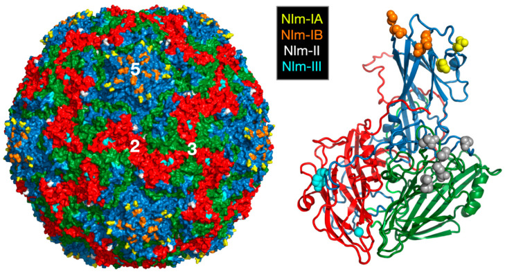
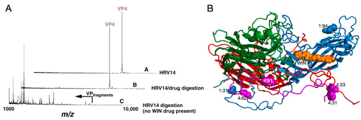
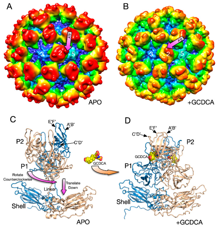
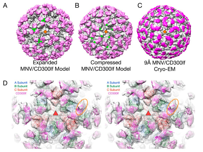
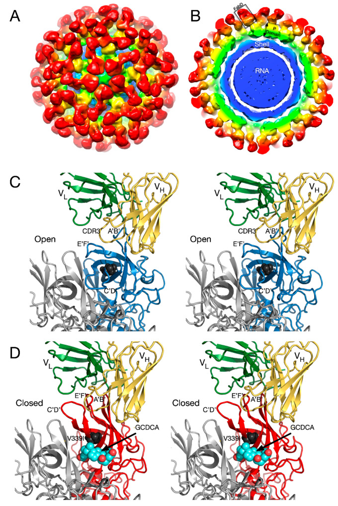
Protein-shelled viruses have been thought as "tin cans" that merely carry the genomic cargo from cell to cell. However, through the years, it has become clear that viruses such as rhinoviruses and caliciviruses are active and dynamic structures waiting for the right environmental cues to deliver their genomic payload to the host cell. In the case of human rhinoviruses, the capsid has empty cavities that decrease the energy required to cause conformational changes, resulting in the capsids "breathing", waiting for the moment when the receptor binds for it to release its genome. Most strikingly, the buried N-termini of VP1 and VP4 are transiently exposed during this process. A more recent example of a "living" protein capsid is mouse norovirus (MNV). This family of viruses have a large protruding (P) domain that is loosely attached to the shell via a single-polypeptide tether. Small molecules found in the gut, such as bile salts, cause the P domains to rotate and collapse onto the shell surface. Concomitantly, bile alters the conformation of the P domain itself from one that binds antibodies to one that recognizes receptors. In this way, MNV appears to use capsid flexibility to present one face to the immune system and a completely different one to attack the host tissue. Therefore, it appears that even protein-shelled viruses have developed an impressive array of tricks to dodge our immune system and efficiently attack the host.

Keywords: antibodies; flexibility; norovirus; rhinovirus.


Conflict of interest statement

The authors declare no conflict of interest.

Figures

Figure 1
Figure 1
Structure of HRV14 and location of the NIm sites. Shown on the left is the surface of a pseudo T = 3 icosahedral capsid. VP1, VP2, and VP3 are shown in blue, green, and red, respectively. The locations of the escape mutation clusters are highlighted as noted. The right figure shows one icosahedral asymmetric unit using the same color scheme.
Figure 2
Figure 2
Use of mass spectrometry to demonstrate HRV14 breathing. (A) MALDI analysis of limited trypsinolysis of HRV14 in the presence and absence of WIN compounds. While HRV14 is extremely sensitive to trypsin cleavage at room temperature, the presence of WIN compounds blocks all cleavage for >18 h. (B) Ribbon diagram of an icosahedra asymmetric unit with VP1, VP2, VP3, and VP4 colored blue, green, red, and mauve, respectively. The locations of trypsin cleavage that occur within the first five minutes of exposure to trypsin are noted by spherical models. Note that there are cleavage sites at the extreme N-termini of both VP1 and VP4 not visible in the crystal structure.
Figure 3
Figure 3
Early cryo-EM image reconstructions of Fab17 (A) and Fab1 (B) bound to HRV14 at ~20Å resolution. (C) Electron density of the 3.5Å crystal structure of the Fab17/HRV14 complex [21]. (D) The ribbon structure of the Fab17/HRV complex. The two Nim-IA escape mutants are highlighted in yellow. (E) Stereo diagram of the electron density of “pocket factor” (grey) in the Fab17/HRV14 crystal structure that came from the PEG400 used for freezing the crystals. For reference, the WIN 52084 drug [10,12] is shown and also moves the protein strand above the density by more than 3.5Å away from the apo conformation (yellow).
Figure 4
Figure 4
Structure of apo MNV. (A) The 8Å cryo-EM structure of MNV colored according to radial distance. (B) Pseudo-atomic model using the atomic model of the P domain [61] and the shell domain [2]. A, B, C subunits are colored blue, green, and red, respectively. The A’B’ and E’F’ loops are colored cyan and tan, respectively. (C) Cross-section of the model from (A) showing the P domain “floating” above the shell domain by more than 18Å. (D) The pseudo-atomic model using the 8Å image reconstruction and the atomic models of the S and P domains. Dimer subunits are colored red and tan, respectively.
Figure 5
Figure 5
Structural changes in the MNV capsid upon the addition of the bile salt, GCDCA. (A) The 8Å structure of apo MNV and (B) the 3Å structure of MNV in the presence of GCDCA and low pass filtered to 8Å for comparison with the apo structure. A and B are colored from blue to red according to radius. (C) The pseudo-atomic model of the apo structure of MNV using the 8Å and 3Å cryo-EM electron density maps. (D) When GCDCA is added, the P domains rotate by ~90° and rest on top of the shell.
Figure 6
Figure 6
The structure of MNV complexed with the receptor, CD300lf. In panels A–C, the virus is grey and the receptor mauve. (A) The model for the expanded form of MNV with the CD300lf place using the atomic structure of the P domain/CD300lf complex [66]. An icosahedral 5-fold axis is represented by an orange pentagon and the C-termini of some of the CD300lf molecules are highlighted with green circles. (B) An atomic model of the MNV/GCDCA/CD300lf complex using the atomic model of the Pdomain/CD300lf and the EM structure of the MNV/GCDCA complex. (C) The 9Å cryo-EM structure of the MNV/GCDCA/CD300lf complex [2]. (D) The model from panel B overlaid with the cryo-EM density of the MNV/GCDCA/CD300lf shown in panel C. An icosahedral 3-fold axis that is denoted by the orange triangle. The orange ellipse notes the CD300lf bound to the A subunit with weaker density likely due to lower occupancy.
Figure 7
Figure 7
Structure of the Fab 2D3.7/MNV complex and possible effects of bile salts on antibody binding. (A) ~9.0Å cryo-EM density of the FabD/MNV complex viewed down an icosahedral 5-fold axis. (B) Central section of the FabD/MNV electron density map. The approximate colors of the shell, P1, and P2 domains are blue, green, and yellow, respectively. The FabD variable domains are orange and the constant domains, red. (B) FabD modeled into the cryo-EM density using the “open” conformation of the P domain. The location of the “allosteric-like” escape to this antibody (V339I) is shown in black. (C) Stereo diagram of the FabD bound to the “open” conformation. (D) Stereo diagram of the hypothetical model of FabD bound to the “closed” conformation induced by the binding of bile salts to illustrate the antibody/P domain clashes in this conformation.

References

    1. Lewis J.K., Bothner B., Smith T.J., Siuzdak G. Antiviral agent blocks breathing of the common cold virus. Proc. Natl. Acad. Sci. USA. 1998;95:6774–6778. doi: 10.1073/pnas.95.12.6774. - DOI - PMC - PubMed
    1. Sherman M.B., Williams A.N., Smith H.Q., Nelson C., Wilen C.B., Fremont D.H., Virgin H.W., Smith T.J. Bile Salts Alter the Mouse Norovirus Capsid Conformation: Possible Implications for Cell Attachment and Immune Evasion. J. Virol. 2019;93:e00970-19. doi: 10.1128/JVI.00970-19. - DOI - PMC - PubMed
    1. Rueckert R.R. Picornaviridae and Their Replication. In: Fields B.N., Knipe D.M., editors. Fundamental Virology. Raven Press; New York, NY, USA: 1996. pp. 609–654.
    1. Rossmann M.G., Arnold E., Erickson J.W., Frankenberger E.A., Griffith J.P., Hecht H.-J., Johnson J.E., Kamer G., Luo M., Mosser A.G., et al. Structure of a human common cold virus and functional relationship to other picornaviruses. Nature. 1985;317:145–153. doi: 10.1038/317145a0. - DOI - PubMed
    1. Colonno R.J., Condra J.H., Mizutani S., Callahan P.L., Davies M.E., Murcko M.A. Evidence for the direct involvement of the rhinovirus canyon in receptor binding. Proc. Natl. Acad. Sci. USA. 1988;85:5449–5453. doi: 10.1073/pnas.85.15.5449. - DOI - PMC - PubMed

Publication types

LinkOut - more resources