Review
. 2020 Jun 5;12(6):1479.
doi: 10.3390/cancers12061479.

Oncogenic Properties of the EBV ZEBRA Protein

Affiliations
Review

Oncogenic Properties of the EBV ZEBRA Protein

Diego Germini et al. Cancers (Basel). .

Abstract

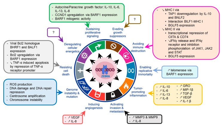
Epstein Barr Virus (EBV) is one of the most common human herpesviruses. After primary infection, it can persist in the host throughout their lifetime in a latent form, from which it can reactivate following specific stimuli. EBV reactivation is triggered by transcriptional transactivator proteins ZEBRA (also known as Z, EB-1, Zta or BZLF1) and RTA (also known as BRLF1). Here we discuss the structural and functional features of ZEBRA, its role in oncogenesis and its possible implication as a prognostic or diagnostic marker. Modulation of host gene expression by ZEBRA can deregulate the immune surveillance, allow the immune escape, and favor tumor progression. It also interacts with host proteins, thereby modifying their functions. ZEBRA is released into the bloodstream by infected cells and can potentially penetrate any cell through its cell-penetrating domain; therefore, it can also change the fate of non-infected cells. The features of ZEBRA described in this review outline its importance in EBV-related malignancies.

Keywords: BZLF1; EBV; ZEBRA; Zta; lytic cycle; oncogenesis; transactivation; transcription; viral-host interaction.


Conflict of interest statement

The authors declare no conflict of interest.

Figures

Figure 1
Figure 1
Epstein-Barr Virus (EBV) life cycle. (1) Infection occurs after the contact with an infected saliva. (2) After initial infection of oropharyngeal epithelial cells, the virus passes into the underlying lymphoid tissue where it infects naive B cells. (3) This immediately triggers the transient pre-latent lytic cycle with expression of ZEBRA and other lytic genes involved in resistance to apoptosis and evasion from the immune system. (4) Infected naive B cells become proliferating B blasts through the growth program (latency III) where all latency proteins are expressed. (5) Cytotoxic T lymphocytes (CTL) trigger a strong immune response (which is impaired during immunodeficiency) to eliminate EBV-infected B cells. (6) Proliferating B blasts migrate into the germinal center (GC) and activate the default transcription program (latency II) where latency protein expression is restricted to EBNA1, LMP1 and LMP2. They differentiate into centroblasts and then centrocytes. (7) Centrocytes leave the GC and differentiate into memory B cells circulating in peripheral blood. These cells have turned off the expression of all viral proteins (latency 0). (8) Occasionally, circulating EBV-positive memory B cells express EBNA1 during homeostatic cell division to ensure viral genome replication and segregation into daughter cells. (9) Following stimulation, latently infected memory B cells can be recruited into GC. (10) Activated EBV-positive memory B cells can differentiate into plasma cells, reactivate the virus and undergo productive lytic cycle that leads to (11) viral shedding into saliva and (12) new naive B cells infection. (13) Activated EBV-positive memory B cells reintegrate the pool of memory B cells. It is not clear whether in vivo stimulated EBV-positive memory B cells which have not differentiated into plasma cells undergo an abortive lytic cycle (ZEBRA and early gene expression without viral production) before reintegrating the pool of memory cells. It is also not clear whether these cells successively re-express different latency programs in the GC in vivo before reintegrating the pool of memory cells.
Figure 2
Figure 2
Structure of the ZEBRA protein. (A) ZEBRA structure. ZEBRA is encoded by the BZLF1 gene containing three exons. ZEBRA protein has an N-terminal transactivation domain (TAD, residues 1-166), a regulatory domain (residues 167–177), a bZIP domain, which consists of a central basic DNA binding domain (DBD, residues 178-194) and a C-terminal coiled-coil dimerization domain (DD, residues 195–221). The minimal domain for cell penetration is located between residues 170-220. Three available partial 3D structures were imported from the SWISS-MODEL Repository [62] (accession number P03206) and are based on crystal structure data published by [39,42,43]. They are shown below the respective primary sequence. Rainbow color code is used to map approximate residue position concordance between primary and tertiary (or quaternary) structure. (B) ZEBRA-response elements (ZREs). Sequences of ZEBRA DNA binding sites (ZREs) of two types: AP-1-like (non-CpG-containing) ZREs and CpG-containing ZREs are depicted as sequence logos, adapted from [51,60].
Figure 3
Figure 3
ZEBRA functions. (A) transcriptional activation by ZEBRA. ZEBRA is shown as a homodimer, relative positions of transactivation domain (TAD), DNA binding domain (DBD) and dimerization domain (DD) are indicated. ZEBRA binds to specific ZEBRA response elements (ZREs) within promoters of viral and host genes with a preference to methylated-CpG DNA. ZEBRA binding leads to sequential recruitment of basal transcription factors and RNA polymerase II. In addition, ZEBRA binds transcriptional coactivator CREB binding protein (CBP). (B) transcriptional repression via ZEBRA binding to cellular transcription factors and by SUMOylated ZEBRA. Transcription factors that interact directly with ZEBRA are listed. The interaction occurs mainly via ZEBRA’s bZIP domain and mutually impedes the function of both ZEBRA and bound transcription factor and results in repression of targeted genes. SUMOylated ZEBRA has a low transactivation activity related to decreased CBP binding and the ability to recruit histone deacetylases (HDAC) to responsive promoters. (C) activation of EBV lytic replication. ZEBRA recognizes the EBV lytic origin (oriLyt), serves as the origin binding protein and recruits viral core replication enzymes to initiate lytic replication of EBV. (D) interaction with cellular proteins not directly involved in transcriptional regulation. ZEBRA interaction partners are listed. ZEBRA interaction with Cul2/Cul5 induces the formation of multimolecular ECS complex (Elongin B/C-Cul2/5-SOCS-box protein) with the ubiquitin ligase activity that targets p53 for proteasomal degradation.
Figure 4
Figure 4
ZEBRA oncogenic properties. ZEBRA directly, or through its target genes, contributes to the acquisition of cancer hallmarks by cells including sustained proliferative signaling, evading or altering the immune response, resisting cell death, enabling replicative immortality, inducing angiogenesis and activating tumor invasion and metastasis. A part of these effects is mediated by genome instability and tumor-promoting inflammation that induce an environment favorable to cancer development and progression. Adapted from [111].

References

    1. Dolcetti R., Dal Col J., Martorelli D., Carbone A., Klein E. Interplay among viral antigens, cellular pathways and tumor microenvironment in the pathogenesis of EBV-driven lymphomas. Semin. Cancer Biol. 2013;23:441–456. doi: 10.1016/j.semcancer.2013.07.005. - DOI - PubMed
    1. Kutok J.L., Wang F. Spectrum of Epstein-Barr Virus–Associated Diseases. Annu. Rev. Pathol. Mech. Dis. 2006;1:375–404. doi: 10.1146/annurev.pathol.1.110304.100209. - DOI - PubMed
    1. Cohen J.I. Epstein-Barr virus infection. N. Engl. J. Med. 2000;343:481–492. doi: 10.1056/NEJM200008173430707. - DOI - PubMed
    1. Tsurumi T., Fujita M., Kudoh A. Latent and lytic Epstein-Barr virus replication strategies. Rev. Med. Virol. 2005;15:3–15. doi: 10.1002/rmv.441. - DOI - PubMed
    1. IARC Proceedings of the IARC Working Group on the Evaluation of Carcinogenic Risks to Humans. Epstein-Barr Virus and Kaposi’s Sarcoma Herpesvirus/Human Herpesvirus 8. Lyon, France, 17-24 June 1997. IARC Monogr. Eval. Carcinog. Risks Hum. 1997;70:1–492. - PubMed

LinkOut - more resources