. 2020 Jun 6;12(6):1484.
doi: 10.3390/cancers12061484.

TGFα Promotes Chemoresistance of Malignant Pleural Mesothelioma

Affiliations

TGFα Promotes Chemoresistance of Malignant Pleural Mesothelioma

Bernard Staumont et al. Cancers (Basel). .

Abstract

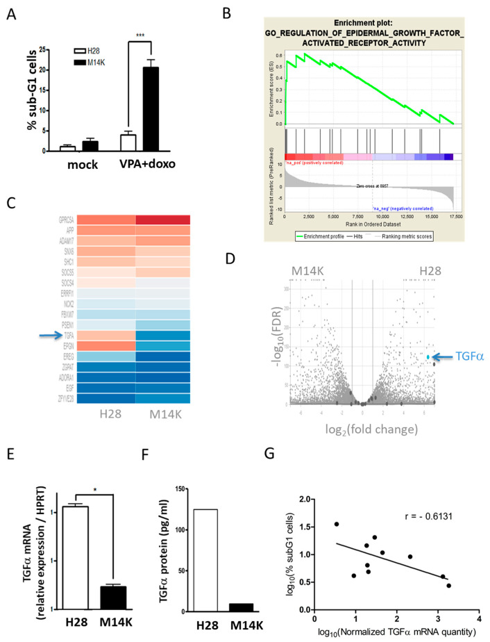
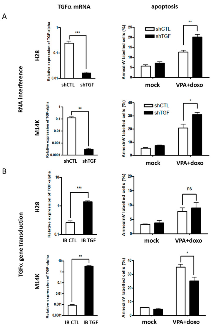
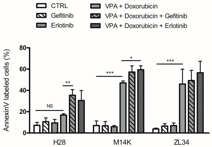
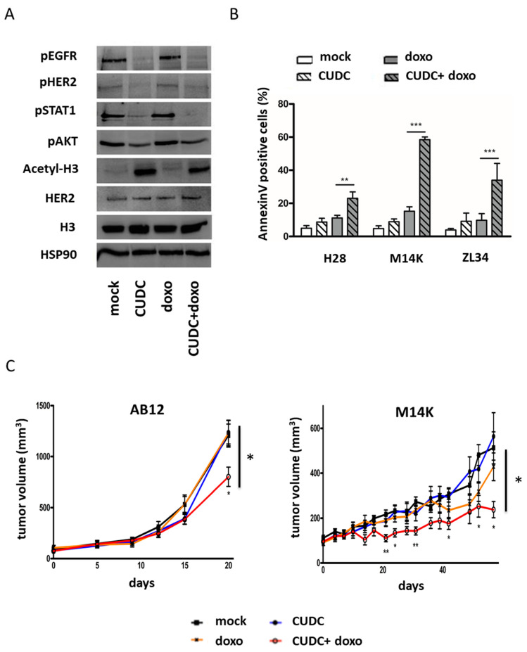
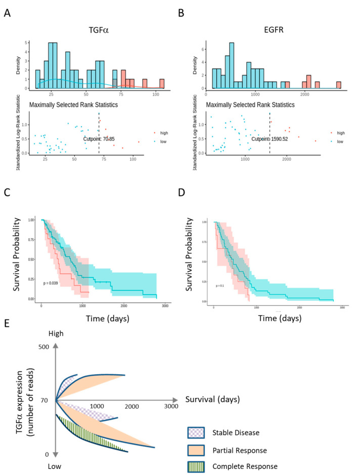
Background: There is no standard chemotherapy for refractory or relapsing malignant pleural mesothelioma (MPM). Our previous reports nevertheless indicated that a combination of an anthracycline (doxorubicin) and a lysine deacetylase inhibitor (valproic acid, VPA) synergize to induce the apoptosis of MPM cells and reduce tumor growth in mouse models. A Phase I/II clinical trial indicated that this regimen is a promising therapeutic option for a proportion of MPM patients. Methods: The transcriptomes of mesothelioma cells were compared after Illumina HiSeq 4000 sequencing. The expression of differentially expressed genes was inhibited by RNA interference. Apoptosis was determined by cell cycle analysis and Annexin V/7-AAD labeling. Protein expression was assessed by immunoblotting. Preclinical efficacy was evaluated in BALB/c and NOD-SCID mice. Results: To understand the mechanisms involved in chemoresistance, the transcriptomes of two MPM cell lines displaying different responses to VPA-doxorubicin were compared. Among the differentially expressed genes, transforming growth factor alpha (TGFα) was associated with resistance to this regimen. The silencing of TGFα by RNA interference correlated with a significant increase in apoptosis, whereas the overexpression of TGFα desensitized MPM cells to the apoptosis induced by VPA and doxorubicin. The multi-targeted inhibition of histone deacetylase (HDAC), HER2 and TGFα receptor (epidermal growth factor receptor/EGFR) improved treatment efficacy in vitro and reduced tumor growth in two MPM mouse models. Finally, TGFα expression but not EGFR correlated with patient survival. Conclusions: Our data show that TGFα but not its receptor EGFR is a key factor in resistance to MPM chemotherapy. This observation may contribute to casting light on the promising but still controversial role of EGFR signaling in MPM therapy.

Keywords: TGFα; chemoresistance; combination therapy; mesothelioma.


Conflict of interest statement

The authors declare no conflict of interest. The funders had no role in the design of the study; in the collection, analyses or interpretation of the data; in the writing of the manuscript; or in the decision to publish the results.

Figures

Figure 1
Figure 1
The sensitivity of mesothelioma (MPM) cells to valproic acid (VPA)-doxorubicin chemotherapy negatively correlates with expression of TGFα. (A) H28 and M14K mesothelioma cells were treated for 24 h with VPA (2 mM) and doxorubicin (100 nM). Apoptotic DNA fragmentation was evaluated by flow cytometry after ethanol fixation and propidium iodide labeling. Data (% of sub-G1 cells) are means ± standard deviations (SD) of three independent experiments. p-value < 0.001 (***) was obtained by the independent Student’s t test. (B) RNA sequencing of H28 and M14K cells. Enrichment plot of epidermal growth factor activated receptor (EGFR) activity gene set determined by gene set enrichment analysis (GSEA). (C) Hierarchical heat map of genes belonging to GO regulation of EGFR activity gene set. (D) Volcano plot based on log2 (fold change) and −log10 (false discovery rate, FDR). (E) The difference in TGFα transcription was confirmed by RT-qPCR in H28 and M14K cells. The HPRT housekeeping gene was used as an endogenous control in order to normalize TGFα expression. RT-qPCR data are represented as means ± SD, and statistical significance was calculated using the Mann-Whitney U test (* p-value < 0.05). (F) Quantification of TGFα protein by ELISA. (G) A correlation test (Pearson) was performed between the log10-transformed data of normalized TGFα expression and the levels of apoptosis induced by VPA-doxorubicin in 10 mesothelioma cell lines (M14K, M38K, MSTO-211H, NCI-H2452, H28, SPC111, SPC212, ZL34, ZL5 and ZL55). Apoptosis was measured by quantifying the proportion of sub-G1 cells by flow cytometry. The results are means of three independent experiments, and the Pearson correlation coefficient (r) is represented on the graph (p-value = 0.0594).
Figure 2
Figure 2
RNA interference and overexpression of TGFα increases and reduces the apoptosis induced by VPA and doxorubicin, respectively. (A) TGFα expression was inhibited in H28 and M14K cells by RNA interference using shRNA cloned into pLKO.1 (shTGF). A scrambled shRNA was used as control (shCTL). Downregulation or increased expression of TGFα was confirmed by RT-qPCR (left panels). The HPRT gene transcript was used as an endogenous control to calculate the relative expression of TGFα. Expression levels are represented as means ± SD of at least three independent experiments. Statistical significance was evaluated with the Mann–Whitney U test. The apoptosis induced by VPA-doxorubicin (VPA + doxo) was measured by an Annexin V assay (right panels). Apoptosis rates were calculated from three independent experiments and are represented as means ± SD. Means were compared using the independent Student’s t-test. * p-value < 0.05; ** p-value < 0.01; *** p-value < 0.001; ns: not significant. (B) TGFα was overexpressed in H28 and M14K cells using the plasmid pCSEF-IB-TGFα (IB TGF), while the empty vector pCSEF-IB served as control (IB CTL). TGFα expression and apoptotic rates were determined as described in panel A.
Figure 3
Figure 3
EGFR tyrosine kinase inhibitors increase the apoptosis induced by VPA and doxorubicin. H28, M14K and ZL34 cells were treated with VPA, doxorubicin and/or an EGFR tyrosine kinase inhibitor (gefitinib or erlotinib) and were evaluated for their apoptotic response by an Annexin V assay. Data are expressed as the means ± SD. Comparisons of multiple groups were performed using one-way analysis of variance (ANOVA) followed by Tukey’s multiple comparison procedure. Data are from three independent experiments. * p-value < 0.05; ** p-value < 0.01; *** p-value < 0.001; NS: not significant.
Figure 4
Figure 4
An inhibitor targeting EGFR/HER2 and HDAC synergizes with doxorubicin to induce apoptosis and inhibit tumor growth. (A) Representative immunoblotting of H28 cell lysates evaluating the phosphorylation of EGFR, STAT1, AKT and HER2 as well as the acetylation of histone H3 in response to doxorubicin and/or the EGFR/HER2/HDAC inhibitor (CUDC-101 at 1 µM). Heat-shock protein 90 (HSP90) was used as a loading control. (B) The apoptosis induced by CUDC-101 (1 µM) and/or doxorubicin (100 nM) was quantified by an Annexin V assay in three mesothelioma cell lines (H28, M14K and ZL34). Data represent the means ± SD of three independent experiments. Statistical significance was determined with one-way analysis of variance (ANOVA) followed by Tukey’s multiple comparisons test. ** p-value < 0.01; *** p-value < 0.001. (C) Tumor growth of AB12 (murine) and M14K (human) mesothelioma cells was evaluated after subcutaneous injection in BALB/c and NOD-SCID mice, respectively. Mice were treated intraperitoneally with mock (30% Captisol), doxorubicin (0.5 mg/kg once a week) or/and CUDC-101 (100 mg/kg twice a week). The tumor volume was measured at regular intervals. One-way analysis of variance (ANOVA) was used at the different time points to evaluate the statistical significance (* p-value < 0.05; ** p-value < 0.01) between the different treatment conditions (six mice per group).
Figure 5
Figure 5
Kaplan-Meier survival curves of patients classified according to TGFα and EGFR expression. TGFα and EGFR expression datasets were downloaded from The Cancer Genome Atlas (TCGA). (A) and (B) Optimal cutpoints between high and low expression levels were calculated by maxstat for TGFα and EGFR, respectively; (C) and (D) Kaplan-Meier survival graphs were generated for patients categorized according to TGFα and EGFR expression; (E) Correlation of therapeutic response with survival of patients characterized by low and high TGFα expression.

References

    1. Yap T.A., Aerts J.G., Popat S., Fennell D.A. Novel insights into mesothelioma biology and implications for therapy. Nat. Rev. Cancer. 2017;17:475–488. doi: 10.1038/nrc.2017.42. - DOI - PubMed
    1. Carbone M., Kanodia S., Chao A., Miller A., Wali A., Weissman D., Adjei A., Baumann F., Boffetta P., Buck B., et al. Consensus report of the 2015 Weinman international conference on mesothelioma. J. Thorac. Oncol. 2016;11:1246–1262. doi: 10.1016/j.jtho.2016.04.028. - DOI - PMC - PubMed
    1. Frost G. The latency period of mesothelioma among a cohort of British asbestos workers (1978–2005) Br. J. Cancer. 2013;109:1965–1973. doi: 10.1038/bjc.2013.514. - DOI - PMC - PubMed
    1. Van den Borre L., Deboosere P. Asbestos in Belgium: An underestimated health risk. The evolution of mesothelioma mortality rates (1969–2009) Int. J. Occup. Environ. Health. 2014;20:134–140. doi: 10.1179/2049396714Y.0000000058. - DOI - PMC - PubMed
    1. Vogelzang N.J., Rusthoven J.J., Symanowski J., Denham C., Kaukel E., Ruffie P., Gatzemeier U., Boyer M., Emri S., Manegold C., et al. Phase III study of pemetrexed in combination with cisplatin versus cisplatin alone in patients with malignant pleural mesothelioma. J. Clin. Oncol. 2003;21:2636–2644. doi: 10.1200/JCO.2003.11.136. - DOI - PubMed