. 2020 Jun 6;12(6):622.
doi: 10.3390/v12060622.

Propagation, Inactivation, and Safety Testing of SARS-CoV-2

Affiliations

Propagation, Inactivation, and Safety Testing of SARS-CoV-2

Alexander S Jureka et al. Viruses. .

Abstract

In late 2019, a novel coronavirus, severe acute respiratory syndrome coronavirus 2 (SARS-CoV-2) emerged in Wuhan, the capital of the Chinese province Hubei. Since then, SARS-CoV-2 has been responsible for a worldwide pandemic resulting in over 4 million infections and over 250,000 deaths. The pandemic has instigated widespread research related to SARS-CoV-2 and the disease that it causes, COVID-19. Research into this new virus will be facilitated by the availability of clearly described and effective procedures that enable the propagation and quantification of infectious virus. As work with the virus is recommended to be performed at biosafety level 3, validated methods to effectively inactivate the virus to enable the safe study of RNA, DNA, and protein from infected cells are also needed. Here, we report methods used to grow SARS-CoV-2 in multiple cell lines and to measure virus infectivity by plaque assay using either agarose or microcrystalline cellulose as an overlay as well as a SARS-CoV-2 specific focus forming assay. We also demonstrate effective inactivation by TRIzol, 10% neutral buffered formalin, beta propiolactone, and heat.

Keywords: SARS-CoV-2; coronavirus; inactivation; plaque assay; virology; virus.


Conflict of interest statement

The authors declare no conflict of interest.

Figures
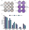
Figure 1
Figure 1
Propagation and quantification of SARS-CoV-2 in cell culture. (A) Vero E6 cells were infected with SARS-CoV-2 at an MOI of 0.001 and monitored daily by microscopy for the presence of cytopathic effect (CPE). (B) Vero E6 cells were infected with serially diluted SARS-CoV-2 stock virus and overlaid with 0.4% agarose in DMEM supplemented with 2% FBS. Plaque assays were harvested at 24, 48, and 72 h post-infection, fixed, and stained with crystal violet to visualize plaques. (C) Vero E6, Calu-3, Caco-2, Huh7, A549, and 293T cells were infected with SARS-CoV-2 at an MOI of 0.01. Supernatants from 24, 48, and 72-h timepoints were quantified by plaque assay. Data are representative of the mean and SEM of 3 replicates.
Figure 2
Figure 2
MCC is a suitable alternative as an overlay medium for SARS-CoV-2 plaque assays. Vero E6 cells were infected with serially diluted SARS-CoV-2 stock virus and overlaid with 0.6% (A) or 1.2% (B) MCC in serum free DMEM. After 72 h, the MCC overlay was removed and monolayer were fixed and stained with crystal violet to visualize plaques.
Figure 3
Figure 3
SARS-CoV-2 focus forming assay. (A) Representative image of the foci observed from serially diluted supernatants from infected Calu-3 cells. (B) Comparison of titers obtained from traditional plaque assay and focus forming assays on matched supernatants collected from SARS-CoV-2 infected Calu-3 cells. Data are representative of the mean and SEM of 3 replicates.
Figure 4
Figure 4
BPL inactivation and purification of SARS-CoV-2 virus particles. (A) Graphical depiction of the workflow established for the inactivation and purification of BPL inactivated SARS-CoV-2. (B) Negative stain transmission electron microscope (TEM) images of SARS-CoV-2 virus particles after purification from cell culture media over a 20% sucrose cushion at 100,000× g for 2 h. Scale bars represent 200 nM. (C) Western blots of BPL inactivated virus particles for SARS-CoV-2 nucleoprotein and spike protein.
Figure 5
Figure 5
Heat Inactivation of SARS-CoV-2. SARS-CoV-2 containing samples (1 × 10⁶ pfu) were heated at 100 °C for 5, 10 and 15 min and 56 °C for 15, 30, 45, and 60 min. Samples were assayed by plaque assay to detect remaining infectious virus post-heating. The room temperature control was incubated at room temperature until all heated samples were prepared. Data are representative of the mean and SEM of 3 replicates.

References

    1. Bedford J., Enria D., Giesecke J., Heymann D.L., Ihekweazu C., Kobinger G., Lane H.C., Memish Z., Oh M.D., Sall A.A., et al. COVID-19: Towards controlling of a pandemic. Lancet. 2020;395:1015–1018. doi: 10.1016/S0140-6736(20)30673-5. - DOI - PMC - PubMed
    1. World Health Organization . Situation Report—112 Coronavirus Disease 2019 (COVID-19) World Health Organization; Geneva, Switzerland: 2020.
    1. Blow J.A., Dohm D.J., Negley D.L., Mores C.N. Virus inactivation by nucleic acid extraction reagents. J. Virol. Methods. 2004;119:195–198. doi: 10.1016/j.jviromet.2004.03.015. - DOI - PubMed
    1. Darnell M.E., Subbarao K., Feinstone S.M., Taylor D.R. Inactivation of the coronavirus that induces severe acute respiratory syndrome, SARS-CoV. J. Virol. Methods. 2004;121:85–91. doi: 10.1016/j.jviromet.2004.06.006. - DOI - PMC - PubMed
    1. Haddock E., Feldmann F., Feldmann H. Effective Chemical Inactivation of Ebola Virus. Emerg. Infect. Dis. 2016;22:1292–1294. doi: 10.3201/eid2207.160233. - DOI - PMC - PubMed

Publication types

MeSH terms