. 2020 Jun 7;9(6):1419.
doi: 10.3390/cells9061419.

Exosome-Derived LINC00960 and LINC02470 Promote the Epithelial-Mesenchymal Transition and Aggressiveness of Bladder Cancer Cells

Affiliations

Exosome-Derived LINC00960 and LINC02470 Promote the Epithelial-Mesenchymal Transition and Aggressiveness of Bladder Cancer Cells

Cheng-Shuo Huang et al. Cells. .

Abstract

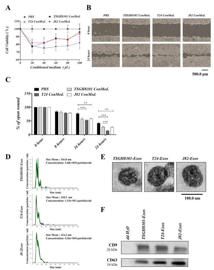
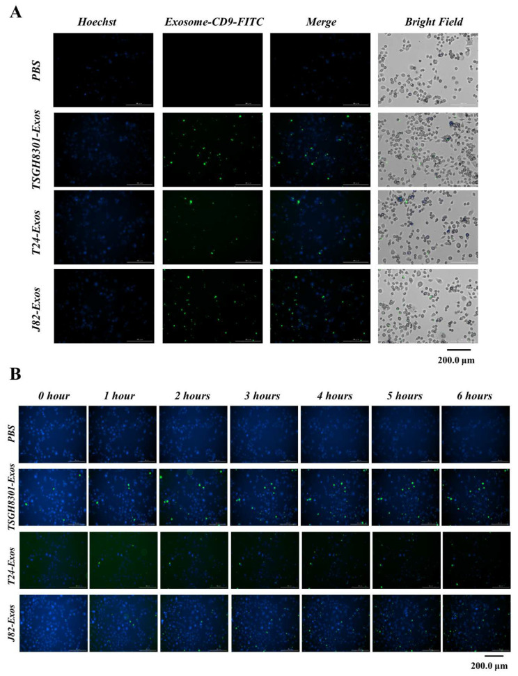
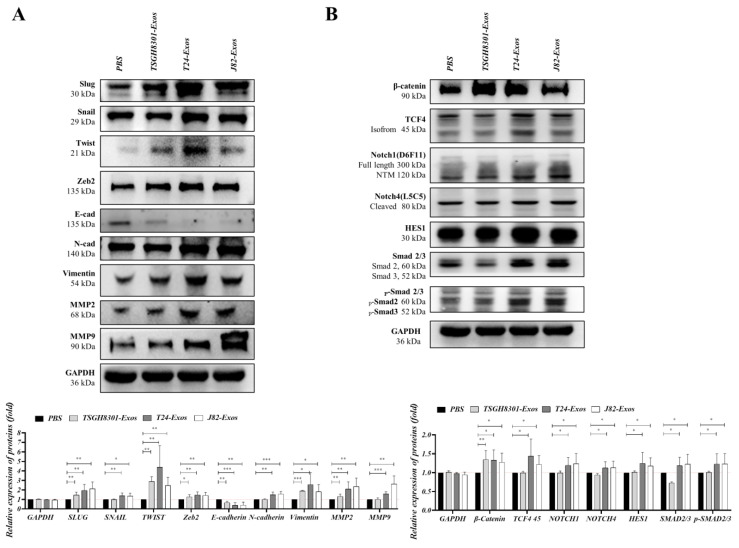
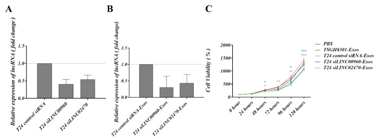
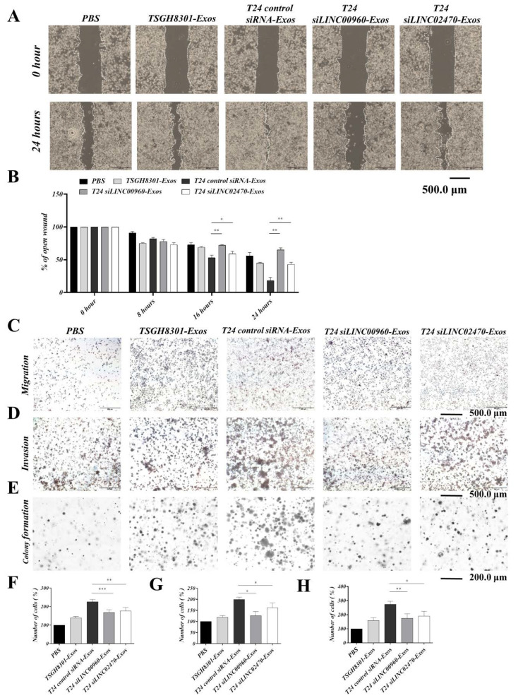
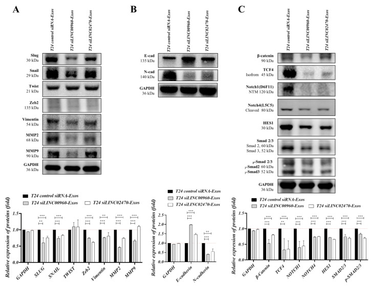
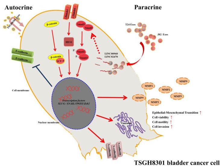
Exosomes are essential for several tumor progression-related processes, including the epithelial-mesenchymal transition (EMT). Long non-coding RNAs (lncRNAs) comprise a major group of exosomal components and regulate the neoplastic development of several cancer types; however, the progressive role of exosomal lncRNAs in bladder cancer have rarely been addressed. In this study, we identified two potential aggressiveness-promoting exosomal lncRNAs, LINC00960 and LINC02470. Exosomes derived from high-grade bladder cancer cells enhanced the viability, migration, invasion and clonogenicity of recipient low-grade bladder cancer cells and activated major EMT-upstream signaling pathways, including β-catenin signaling, Notch signaling, and Smad2/3 signaling pathways. Nevertheless, LINC00960 and LINC02470 were expressed at significantly higher levels in T24 and J82 cells and their secreted exosomes than in TSGH-8301 cells. Moreover, exosomes derived from LINC00960 knockdown or LINC02470 knockdown T24 cells significantly attenuated the ability of exosomes to promote cell aggressiveness and activate EMT-related signaling pathways in recipient TSGH-8301 cells. Our findings indicate that exosome-derived LINC00960 and LINC02470 from high-grade bladder cancer cells promote the malignant behaviors of recipient low-grade bladder cancer cells and induce EMT by upregulating β-catenin signaling, Notch signaling, and Smad2/3 signaling. Both lncRNAs may serve as potential liquid biomarkers for the prognostic surveillance of bladder cancer progression.

Keywords: bladder cancer; epithelial-mesenchymal transition; exosome; long non-coding RNAs.


Conflict of interest statement

The authors declare no conflict of interest.

Figures

Figure 1
Figure 1
Conditioned medium of high-grade bladder cancer cells increased viability and motility of low-grade bladder cancer cells. (A) Cell viability was compared using MTT assays in TSGH-8301 cells treated with the indicated conditioned medium, ** p < 0.01, *** p < 0.001. (B,C) The wound healing assay demonstrated that conditioned medium increased the migration of TSGH-8301 cells. Wound areas were measured at 0, 8, 16, and 24 h after scratching, and the representative images were shown at 0 h and 24 h after scratching. The wound closure distance was measured with the ImageJ software. The bars represent the mean and SD of three independent experiments, ** p < 0.01, *** p < 0.001. (D) Nanoparticle tracking analysis was used to compare the average size of isolated exosomes. (E) Transmission electron microcopy was used to observe the morphology of isolated exosome. (F) Western blots revealed the presence of exosomal markers, CD9 and CD63, in isolated exosomes.
Figure 2
Figure 2
Exosomes serve as a mediator in intercellular communication. Exosomes derived from bladder cancer cells were labeled with FITC-conjugated anti-CD9 antibody (green) and counterstained with Hoechst 33342 (blue) for 24 h, and exosomes uptake was observed under a BioTek LIONheart FX microscope (A) for different spectra, and (B) merged figures were aligned upon different time points.
Figure 3
Figure 3
T24-Exos and J82-Exos intensified the aggressive behavior of TSGH-8301 cells. (A) Cell viability of different concentrations and (B) cell growth curve of different time points were compared using MTT assays in TSGH-8301 cells treated with each exosome, * p < 0.05, ** p < 0.01, *** p < 0.001. (C,D) The wound healing assay demonstrated that T24 and J82 cell-derived exosomes increased the migratory abilities of TSGH-8301 cells. Wound areas were photographed at 0, 8, 16, and 24 h after scratching, and representative images are shown at 0 h and 24 h after scratching. The wound closure distance was calculated with the ImageJ software. The bars represent the mean and SD of three independent experiments, * p < 0.05, ** p < 0.01, *** p < 0.001. (E,H) Transwell assay showed that T24 and J82 cell-derived exosomes increased the migratory abilities of TSGH-8301 cells. (F,I) Matrigel-coated transwell assay showed that T24 and J82 cells derived exosomes increased the invasive abilities of TSGH-8301 cells. Representative images of the (E) migrated and (F) invaded cells were photographed at 24 and 72 h after cell inoculation, respectively (G,J) Soft-agar colony formation assay was performed to examine the anchorage-independent survival of cells. The bar-charts showed the quantitative change in (H) migration, (I) invasion, and (J) cell clones, respectively. All experiments were performed at least in triplicate, * p < 0.05, ** p < 0.01, *** p < 0.001.
Figure 4
Figure 4
Exosomes mediated the expression of epithelial–mesenchymal transition-related molecules. (A) Protein expression levels of EMT effectors: Slug, Snail, Twist, Zeb2, E-Cadherin, N-Cadherin, vimentin, MMP2, and MMP9, and (B) EMT-induced signaling molecules: β-catenin, TCF4, Notch1, Notch4, HES1, Smad2/3, and p-Smad2/3 were compared after treatment with exosomes. The bar-charts showed the mean and SD of triplicate experiments, * p < 0.05, ** p < 0.01, *** p < 0.001.
Figure 5
Figure 5
Screening and validation of lncRNA candidates. (A) Schematic diagram of lncRNA screening. Seventeen candidate lncRNAs were screened from the intersection between the top 200 lncRNA of normal vs. tumor and low-grade vs. high-grade bladder cancer datasets in the lnCAR database. (B) Intracellular expression levels of AATBC, LINC00958, LINC00960, SNHG18, MIR4697HG, MEG3, TRMU, IGFL2-AS1, LINC01451, GLIDR, LOC728673, LINC01637, LINC01291, LINC02740, XIST, KLF3-AS1, SSTR5-AS1 were among the different bladder cancer cells that were compared using RT-qPCR. (C) Exosomal levels of LINC00958, LINC00960, MIR4697HG, LINC01637, LINC01291, LINC02740 and XIST were also compared using RT-qPCR. (D) Exosome-derived LINC00960 or LINC02470 transferred into TSGH-8301 was also evaluated by RT-qPCR. The bar-charts showed the mean and SD of triplicate experiments, * p < 0.05, ** p < 0.01, *** p < 0.001.
Figure 6
Figure 6
Exosomes derived from LINC00960-knockdown or LINC02470-knockdown T24 cell conferred less aggressiveness-inductive abilities in recipient TSGH-8301 cells. LINC00960 or LINC02470 expression levels were compared by RT-qPCR in (A) intracellular levels and (B) the exosomal levels. (C) Cell growth curve was compared using MTT assays in TSGH-8301 cell treated with each exosome. The quantitative results are shown as the bar-charts or growth curves for mean and SD of triplicate experiments, * p < 0.05, ** p < 0.01, *** p < 0.001.
Figure 7
Figure 7
Exosomes derived from LINC00960-knockdown or LINC02470-knockdown T24 cell conferred less aggressiveness-inductive abilities in recipient TSGH-8301 cells. T24 siLINC00960-Exos or T24 siLINC02470-Exos induced less migratory ability (A,B for wound healing assay and C,F for transwell assay), invasive ability (D,G for Matrigel-coated transwell assay), and clonogenicity (E,H for soft-agar colony formation assay) of recipient TSGH-8301 cell when compared to T24-Exos. All experiments were performed with the identical time-points shown in Figure 3, and the bar-charts showed the mean and SD of triplicate experiments, * p < 0.05, ** p < 0.01, *** p < 0.001.
Figure 8
Figure 8
Exosomes derived from LINC00960-knockdown or LINC02470-knockdown T24 cell reduced the expression levels of EMT-related molecules in recipient TSGH-8301 cells. (A) Protein expression levels of EMT effectors: Snail, Slug, Twist, Zeb2, vimentin, MMP2, MMP9, (B) E-Cadherin and N-Cadherin, and (C) EMT-induced signaling molecules: β-catenin, TCF4, Notch1, Notch4, HES1, Smad2/3 and p-Smad2/3 were compared after treatment with each exosome. The bar-charts showed the mean and SD of triplicate experiments, ** p < 0.01, *** p < 0.001.
Figure 9
Figure 9
Schematic diagram of the LINC00960 and LINC02470-based signaling circuit in low-grade bladder cancer. T24-Exos and J82-Exos-derived LINC00960 and LINC02470 increased the expression of β-catenin signaling, Notch signaling, Smad2/3 signaling, transcription factors Snail, Slug, N-Cadherin, vimentin, MMP2, and MMP9, decreased E-Cadherin and promoted tumor progression of recipient TSGH-8301 cells.

References

    1. Wong M.C.S., Fung F.D.H., Leung C., Cheung W.W.L., Goggins W.B., Leung A.K. The global epidemiology of bladder cancer: A joinpoint regression analysis of its incidence and mortality trends and projection. Sci. Rep. 2018;8:1129. doi: 10.1038/s41598-018-19199-z. - DOI - PMC - PubMed
    1. Ferlay J., Ervik M., Lam F., Colombet M., Mery L., Piñeros M., Znaor A., Soerjomataram I., Bray F. Global Cancer Observatory: Cancer Tomorrow. WHO; Geneva, Switzerland: 2018.
    1. Bureau of Health Promotion, Department of Health, the Executive Yuan, Taiwan: Cancer Registry Annual Report, 2012, Taiwan. 27. Health Promotion Administration, Ministry of Health and Welfare, Taiwan. Taiwan Cancer Registry Annual Report. [(accessed on 22 July 2019)]; Available online: https://www.hpa.gov.tw/Pages/List.aspx?nodeid=269.
    1. Van Rhijn B., Burger M., Lotan Y., Solsona E., Stief C.G., Sylvester R.J., Witjes J.A., Zlotta A.R. Recurrence and Progression of Disease in Non–Muscle-Invasive Bladder Cancer: From Epidemiology to Treatment Strategy. Eur. Urol. 2009;56:430–442. doi: 10.1016/j.eururo.2009.06.028. - DOI - PubMed
    1. Babjuk M. Bladder Cancer in the Elderly. Eur. Urol. 2017;73:51–52. doi: 10.1016/j.eururo.2017.04.018. - DOI - PubMed

Publication types

MeSH terms

Substances