. 2020 Jun 10;287(1928):20192918.
doi: 10.1098/rspb.2019.2918. Epub 2020 Jun 10.

Evidence that eye-facing photophores serve as a reference for counterillumination in an order of deep-sea fishes

Affiliations

Evidence that eye-facing photophores serve as a reference for counterillumination in an order of deep-sea fishes

Alexander L Davis et al. Proc Biol Sci. .

Abstract

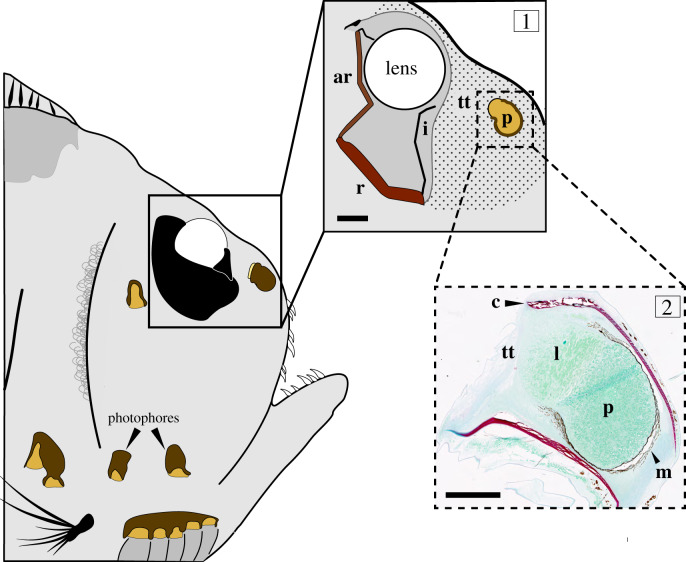
Counterillumination, the masking of an animal's silhouette with ventral photophores, is found in a number of mesopelagic taxa but is difficult to employ because it requires that the animal match the intensity of downwelling light without seeing its own ventral photophores. It has been proposed that the myctophid, Tarletonbeania crenularis, uses a photophore directed towards the eye, termed an eye-facing photophore, as a reference standard that it adjusts to match downwelling light. The potential use of this mechanism, however, has not been evaluated in other fishes. Here, we use micro-computed tomography, photography and dissection to evaluate the presence/absence of eye-facing photophores in three families of stomiiform fishes. We found that all sampled species with ventral photophores capable of counterillumination possess an eye-facing photophore that is pigmented on the anterior and lateral sides, thus preventing its use as a laterally directed signal, lure or searchlight. The two species that are incapable of counterillumination, Cyclothone obscura and Sigmops bathyphilus, lack an eye-facing photophore. After determining the phylogenetic distribution of eye-facing photophores, we used histology to examine the morphology of the cranial tissue in Argyropelecus aculeatus and determined that light from the eye-facing photophore passes through a transparent layer of tissue, then the lens, and finally strikes the accessory retina. Additionally, eight of the 14 species for which fresh specimens were available had an aphakic gap that aligned with the path of emitted light from the eye-facing photophore, while the remaining six had no aphakic gap. These findings, combined with records of eye-facing photophores from distantly related taxa, strongly suggest that eye-facing photophores serve as a reference for counterillumination in these fishes.

Keywords: bioluminescence; camouflage; deep-sea; stomiiformes.


Conflict of interest statement

Authors declare no competing interests.

Figures

Figure 1.
Figure 1.
Micro-CT images of (a) Sternoptyx pseudobscura, (b) Argyropelecus hemigymnus and (c) Sternoptyx diaphana showing the presence and orientation of the photophores from iodine-stained specimens. The eye-facing photophore and the lens are shown in the inset panels. Eye-facing photophores and ventral photophores denoted by the arrows and brackets, respectively. Top: lateral view. Bottom: ventral view. All scale bars are 5 mm. (Online version in colour.)
Figure 2.
Figure 2.
Light path from eye-facing photophore of three representative species. Micro-CT images from (a) A. hemigymnus, (b) S. diaphana and (c) M. weitzmani, showing zoomed-in side and top views of the eye-facing photophore and the eye. The dashed lines for all views are parallel to face of the photophore closest to the eye. Arrows are perpendicular to the dashed lines and denote the predicted light path. The top view for S. diaphana is in the plane of the dorsal surface of the photophore, not in the plane of the most-dorsal surface of the body, because the photophore is obscured dorsally by the musculature (note––the musculature does not extend into the light path between the photophore and the eye). (Online version in colour.)
Figure 3.
Figure 3.
(a) Phylogeny of sampled species of Gonostomatidae, Sternoptychidae and Phosichthyidae pruned from a comprehensive molecular phylogeny of fishes [15]. Colours indicate the presence or absence of the ventral photophores, the eye-facing photophore and an aphakic gap (where available). Only 25/36 species are included in the tree, but the remaining 11 species all exhibit ventral photophores capable of counterillumination and eye-facing photophores (Ariaophos eastropas, Agyripnus atlanticus, A. brocki, A. ephippiatus, Argyropelecus pacificus, Cyclothone braueri, Maurolicus muelleri, Polyipnus aquavitus, P. nuttingi, P. spinifer, Vinciguerria poweriae). Dissecting scope images show examples of the eye-facing photophore surrounded by melanin in (b) Cyclothone pallida, (c) Argyropelecus lychnus and (d) Maurolicus japonicus. The eye-facing photophores are denoted by the white arrows. (Online version in colour.)
Figure 4.
Figure 4.
Digital illustration of Argyropelecus aculeatus showing the morphology of the eye and eye-facing photophore constructed from micro-CT, histological measurements, and previously described retinal morphology [27]. Box 1: the eye-facing photophore (p) emits light that passes through a volume of transparent tissue (tt) and through a dip in the iris (i) reported by Collin et al. [27]. After travelling through the lens, light strikes the accessory retina (ar) while downwelling light illuminates the main retina (r). Box 2: histological section through the middle of the eye-facing photophore shows photocytes (p), the photophore lens (l) and a layer of melanin (m) surrounding three sides of the photophore, preventing light from escaping anteriorly or laterally. Note, there is no reflector or filter in the eye-facing photophore. Additionally, collagen layers (c) flare out from the end of the photophore that the light exits. Once leaving the photophore, light travels through tissue (tt) that is largely transparent and does not stain with Fast Green. Scale bars: box 1, 500 µm; box 2, 100 µm. (Online version in colour.)

References

    1. Johnsen S. 2014. Hide and seek in the open sea: pelagic camouflage and visual countermeasures. Ann. Rev. Mar. Sci. 6, 369–392. (10.1146/annurev-marine-010213-135018) - DOI - PubMed
    1. Claes JM, Mallefet J. 2010. The lantern shark's light switch: turning shallow water crypsis into midwater camouflage. Biol. Lett. 6, 685–687. (10.1098/rsbl.2010.0167) - DOI - PMC - PubMed
    1. Clarke WD. 1963. Function of bioluminescence in mesopelagic organisms. Nature 198, 1244–1246. (10.1038/1981244a0) - DOI
    1. Herring PJ. 1977. Bioluminescence of marine organisms. Nature 267, 788–793. (10.1038/267788a0) - DOI
    1. Widder EA. 1999. Bioluminescence. In Adaptive mechanisms in the ecology of vision (eds Archer SN, Djamgoz MBA, Loew ER, Partridge JC, Vallerga S), pp. 555–581. Boston, MA: Kluwer Academic.

Publication types