. 2020 Jul 31;295(31):10794-10806.
doi: 10.1074/jbc.RA120.013939. Epub 2020 Jun 9.

The J-elongated conformation of β2-glycoprotein I predominates in solution: implications for our understanding of antiphospholipid syndrome

Affiliations

The J-elongated conformation of β2-glycoprotein I predominates in solution: implications for our understanding of antiphospholipid syndrome

Eliza Ruben et al. J Biol Chem. .

Abstract

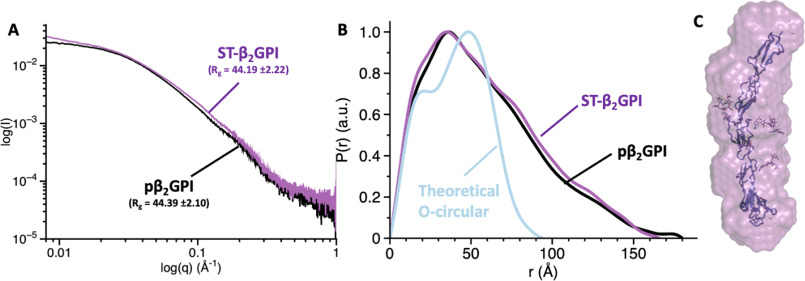
β2-Glycoprotein I (β2GPI) is an abundant plasma protein displaying phospholipid-binding properties. Because it binds phospholipids, it is a target of antiphospholipid antibodies (aPLs) in antiphospholipid syndrome (APS), a life-threatening autoimmune thrombotic disease. Indeed, aPLs prefer membrane-bound β2GPI to that in solution. β2GPI exists in two almost equally populated redox states: oxidized, in which all the disulfide bonds are formed, and reduced, in which one or more disulfide bonds are broken. Furthermore, β2GPI can adopt multiple conformations (i.e. J-elongated, S-twisted, and O-circular). While strong evidence indicates that the J-form is the structure bound to aPLs, which conformation exists and predominates in solution remains controversial, and so is the conformational pathway leading to the bound state. Here, we report that human recombinant β2GPI purified under native conditions is oxidized. Moreover, under physiological pH and salt concentrations, this oxidized form adopts a J-elongated, flexible conformation, not circular or twisted, in which the N-terminal domain I (DI) and the C-terminal domain V (DV) are exposed to the solvent. Consistent with this model, binding kinetics and mutagenesis experiments revealed that in solution the J-form interacts with negatively charged liposomes and with MBB2, a monoclonal anti-DI antibody that recapitulates most of the features of pathogenic aPLs. We conclude that the preferential binding of aPLs to phospholipid-bound β2GPI arises from the ability of its preexisting J-form to accumulate on the membranes, thereby offering an ideal environment for aPL binding. We propose that targeting the J-form of β2GPI provides a strategy to block pathogenic aPLs in APS.

Keywords: X-ray crystallography; antiphospholipid syndrome; autoimmune disease; autoimmunity; beta-2 glycoprotein I; coagulation; complement system; lipid–protein interaction; protein–protein interaction; single-molecule biophysics; structural biology; structure-function; thrombosis.


Conflict of interest statement

Conflict of interest—The authors declare that they have no conflicts of interest with the contents of this article.

Figures

Figure 1.
Figure 1.
Structure of human β2GPI and current mechanism of antigen-antibody recognition for pathogenic anti-DI antibodies in APS. A, color-coded domain structure of human β2GPI (pβ2GPI). β2GPI consists of 326 amino acids organized into five domains (DI–V) connected by four short linkers, Lnk1 (residues 61–64), Lnk2 (residues 119–122), Lnk3 (residues 182–185), and Lnk4 (residues 242–244) (3). Domains I–IV are canonical complement control protein (CCP) domains, each containing two disulfide bonds. In contrast, DV is aberrant, consisting of one extra disulfide bond and a 19-residue hydrophobic loop that is responsible for anchoring the protein to negatively charged phospholipids (7, 8). Similar to other CCP-containing proteins, β2GPI is also heavily glycosylated, bearing four N-linked and one O-linked glycosylations located at positions T130, N143, N164, N174, and N234 that account for ∼20% of the total protein mass. The position of the O- and N-linked glycosylations is shown as triangles and stars, respectively. B, based on previous studies, β2GPI is believed to adopt an O-circular (29, 31), an S-twisted (32), and a J-elongated conformation (33, 34). The J-open conformation results upon interaction of DV with the phospholipids exposing the cryptic epitope R39-R43 (purple) to the solvent. The O-circular form features an intramolecular interaction between DI (blue) and DV (yellow), with amino acids K19, R39, and R43 in DI potentially making contact with K305 and K317 in DV. The S-twisted conformation features a rotation of the DI/DII module, brokered by Lnk2, relative to the rest of the protein, resulting in the blockade of the immunogenic region R39-R43 by the N-linked glycosylation (orange line).
Figure 2.
Figure 2.
Functional characterization of human recombinant β2GPI. A, color-coded domain structure of the recombinant variants used in this work (i.e. LT-β2GPI, hrβ2GPI, and ST-β2GPI), highlighting the position and chemical composition of the N-terminal tag. The long tag (LT, yellow) is composed of three parts: (i) a calcium-dependent epitope for the mAb HPC4 (EDQVDPRLIDGK); (ii) a site-specific biotinylation sequence (AviTag); and (iii) a conventional enterokinase recognition site (DDDDK). Two flexible linkers (i.e. GGGS) were introduced to separate the three functional units of the tag to avoid the formation of secondary structure and ensure exposure of the tag to solvent. Removal of the LT with enterokinase generates hrβ2GPI. The short-tag version of β2GPI contains only the HPC4 purification tag (purple). B, SDS-PAGE analysis of the recombinant proteins (sample 1, LT-β2GPI; sample 2, hrβ2GPI; sample 3, ST-β2GPI) and plasma-purified β2GPI (sample 4) before (left) and after (right) the removal of the glycosylations. Protein deglycosylation mix II, from NEB, was used to remove O-linked and N-linked glycosylations under denaturing conditions. Chemical identity was verified by N-terminal sequencing, and results are the following: band 1, EDQVD; band 2, GRT; band 3, EDQVD; band 4, GRTC. C, representative sensograms of the interaction between LT-β2GPI and liposomes (PC:PS, 80:20) monitored by SPR. Liposomes were immobilized on an L1 chip and soluble β2GPI (0–2 μm) was used in the fluid phase. D, dose-dependent curves quantifying the interaction of LT-β2GPI (red circles), hrβ2GPI (blue circles), ST-β2GPI (green circles), and pβ2GPI (gray circles) with liposomes monitored by SPR. Affinity values (Kd) are 0.19 ± 0.05 μm for LT-β2GPI, 0.33 ± 0.08 μm for hrβ2GPI, 0.22 ± 0.08 μm for ST-β2GPI, and 0.32 ± 0.09 μm for pβ2GPI. No significant binding was observed with liposomes made entirely of PC (light gray circles). Each experiment was repeated at least three times, using two distinct batches of proteins. E, reactivity of immobilized LT-β2GPI (red bars), hrβ2GPI (blue bars), ST-β2GPI (green bars), and pβ2GPI (gray bars) against IgG anti-β2GPI antibodies followed by ELISA. Comparisons between 2 groups were performed using a two-sample t test. Results were considered significant at p < 0.05 (*).
Figure 3.
Figure 3.
X-ray crystal structures of human recombinant β2GPI. X-ray crystal structures of hrβ2GPI (blue) (A), ST-β2GPI (green) (B), and pβ2GPI (gray) (C) solved at 2.6-, 3.0-, and 2.4-Å resolution. All three structures document similar elongated conformations of the protein spanning ∼140 Å in length. Disulfide bonds are highlighted in yellow. D, zoom-in of three extra residues (DGK) belonging to the N-terminal tag preceding the natural sequence of β2GPI (1GRTC4) exclusively found in the structure of ST-β2GPI, as expected. The electron density 2F0-Fc map is countered at 1.0σ. E, structural architecture of the epitope R39-R43 in DI highlighting the position and interactions of R43 (magenta stick) with the nearby residues R39, G41, and T57. Hydrogen bonds between the guanidinium group of R43 and neighboring residues are shown in black. Of note, the conformation of this segment is not involved in crystal contacts and is conserved in all the available crystal structures of β2GPI solved thus far, despite the high salt concentrations in which the crystals grow, suggesting that this is a genuine structural feature of β2-GPI.
Figure 4.
Figure 4.
Location and structural role of the N-linked glycosylations. A, superposition of five X-ray crystal structures of β2GPI highlights structural similarities yet diversity of the phospholipid binding loop in DV (residues 308–319). B, extra electron density detected in the structure of pβ2GPI solved at 2.4-Å resolution attributed to the N-linked glycosylations. The domains of β2GPI are color coded as shown in Fig. 1. The presence of a putative O-linked glycosylation at T130 could not be confirmed because of weak density. Side (C) and top (D) views of the N-glycosylations surrounding DIII are shown. The electron density 2F0-Fc map is countered at 0.8σ.
Figure 5.
Figure 5.
smFRET measurements of β2GPI in solution. A, confocal microscope used to collect smFRET data of fluorescently labeled β2GPI showing the ps-pulsed laser box, the 60× objective, the observation chamber (confocal volume), the location of the pinhole, and two SPAD detectors. B, structure-based design of the FRET constructs S13C/S112C, S13C/S312C, S112C/S312C, and S190C/S312C used in this study. Ser residues mutated to Cys for conjugation with the thiol-reactive dyes AF555 and AF647 used in smFRET measurements are indicated by red spheres. C, FRET couples are listed with their respective domains. Cα-Cα distances were obtained from the crystal structure of hrβ2GPI. D, after labeling and gel filtration, selective incorporation of the fluorescence dyes was verified by loading the proteins (samples 2–5) alongside β2GPI WT (sample 1) into a gradient 4–12% polyacrylamide gel in the presence of SDS and visualized by Coomassie brilliant blue R-250 (black and white) or fluorescence intensity by exciting donor at 532 nm (red panel) and acceptor at 640 nm (blue panel). The uncut gel is shown in Fig. S2C. smFRET histograms of the mutants S13C/S312C, S112C/S312C, S13C/S112C, and S190C/S312C were labeled with AF555/647 measured in Tris 20 mm, pH 7.4, 145 mm NaCl, 0.1% Tween 20 for 1 h at room temperature at a concentration of 100 pm. Populations were fit to a single Gaussian distribution (black lines). FRET efficiency values and calculated distances are indicated. The theoretical shot noise peak highlighting conformational heterogeneity for the FRET pair 190/312 is shown as a dotted line. F, effect of sodium chloride monitored by smFRET. smFRET experiments for the mutant 13/312 (100 pm) were performed at 25, 145, and 1000 mm NaCl in 20 mm Tris, pH 7.4, 145 mm NaCl. G, effect of pH monitored by smFRET. smFRET experiments for the mutant 13/312 (100 pm) were performed at pH 4.0 (20 mm Na acetate, pH 4.0, 150 mm NaCl), pH 7.4 (20 mm Tris, pH 7.4, 145 mm NaCl), and pH 10.0 (20 mm glycine, pH 10.0, 145 mm NaCl).
Figure 6.
Figure 6.
Elongated conformation of β2GPI revealed by SAXS. Scattering profiles (A) and pair distribution functions (B) for pβ2GPI (black) and ST-β2GPI (magenta) collected at 2 mg/ml under physiological conditions (20 mm Tris, pH 7.4, 145 mm NaCl). The calculated values of the radius of gyration, Rg, are very similar for pβ2GPI and ST-β2GPI. The blue curve in panel B, which is significantly different from the experimental scattering profiles, represents the theoretical pair distribution function for a hypothetical circular conformation. C, ab initio envelope calculated from scattering profiles for ST-β2GPI (magenta). Note that the linear arrangement of domains in the structure 6V09 is consistent with the elongated SAXS envelope.
Figure 7.
Figure 7.
Exposure of DI revealed by MBB2 binding and mutagenesis studies. A, interaction of MBB2 and LT-β2GPI monitored by SPR. MBB2 was immobilized onto a CM5 chip using NHS/EDC chemistry to a final density of 6000 RU. A solution of LT-β2GPI (0–20 μm) in Tris 20 mm, pH 7.4, 145 mm NaCl, 0.01% Tween 20 was injected at 30 µl/min for 60 s to observe binding, followed by 60 s of dissociation in running buffer. B, dose-dependent curves quantifying the interaction of LT-β2GPI (red circles), hrβ2GPI (blue circles), ST-β2GPI (green circles), and pβ2GPI (gray circles) with MBB2 monitored by SPR. Affinity values (Kd) are 2.2 ± 0.5 μm for LT-β2GPI, 2.1 ± 0.6 μm for hrβ2GPI, 1.8 ± 0.5 μm for ST-β2GPI, and 1.4 ± 0.6 μm for pβ2GPI. Each experiment was repeated at least three times, using two distinct batches of proteins. C, effect of sodium chloride. SPR binding experiments were performed at 15, 75, 150, and 300 mm NaCl. Analysis of the slope of the linear fit of the association constant versus [Na+] reveals a strong dependence of Na+ and the presence of at least 2.0 ionic contacts (salt bridges) in the complex (59, 60). D, electrostatic potential displaying positive (blue) and negative (red) clusters with a −2.0 to 2.0 intensity scale. The N-glycosylations are shown as magenta sticks. Locations of residues R39, R43, K44, and 120–122 were targeted by site-directed mutagenesis. E, SPR analysis of β2GPI WT and mutant R39A/R43A/RK44A reveals that residues R39 and R43 are critical for MBB2 binding. F, SPR analysis of β2GPI WT (black circles), DI (green circles), β2GPIΔ(120-122) (magenta circles), and deglycosylated β2GPI (blue circles). Removal of DII–DV, perturbation of Lnk2, and removal of the glycosylations do not affect the binding affinity of MBB2 toward β2GPI. This indicates that 1) DI is primed for autoantibody recognition in solution, 2) the exposure of residues R39 and R43 in DI is independent of the conformation of Lnk2, and 3) Lnk2 is not an epitope of MBB2. Given the different molecular weights of the constructs, the binding curves are reported as normalized fraction bound versus analyte to facilitate comparison. Each experiment was repeated at least three times, using two distinct batches of proteins.

References

    1. Schultze H. E., Heide K., and Haupt H. (1961) Uber ein bischer unb ekanntesniedermolekularis b2-globulins des human serums. Naturwissens-chaften 48, 719–719 10.1007/BF00620967 - DOI
    1. de Groot P. G., and Meijers J. C. (2011) Beta(2) glycoprotein I: evolution, structure and function. J. Thromb. Haemost. 9, 1275–1284 10.1111/j.1538-7836.2011.04327.x - DOI - PubMed
    1. Lozier J., Takahashi N., and Putnam F. W. (1984) Complete amino acid sequence of human plasma beta 2-glycoprotein I. Proc. Natl. Acad. Sci. U S A 81, 3640–3644 10.1073/pnas.81.12.3640 - DOI - PMC - PubMed
    1. Lehtinen M. J., Meri S., and Jokiranta T. S. (2004) Interdomain contact regions and angles between adjacent short consensus repeat domains. J. Mol. Biol. 344, 1385–1396 10.1016/j.jmb.2004.10.017 - DOI - PubMed
    1. Forneris F., Wu J., Xue X., Ricklin D., Lin Z., Sfyroera G., Tzekou A., Volokhina E., Granneman J. C., Hauhart R., Bertram P., Liszewski M. K., Atkinson J. P., Lambris J. D., and Gros P. (2016) Regulators of complement activity mediate inhibitory mechanisms through a common C3b-binding mode. EMBO J. 35, 1133–1149 10.15252/embj.201593673 - DOI - PMC - PubMed

Publication types

Substances

Associated data