. 2020 Jun 9;11(1):2911.
doi: 10.1038/s41467-020-16665-z.

ADP binding by the Culex quinquefasciatus mosquito D7 salivary protein enhances blood feeding on mammals

Affiliations

ADP binding by the Culex quinquefasciatus mosquito D7 salivary protein enhances blood feeding on mammals

Ines Martin-Martin et al. Nat Commun. .

Abstract

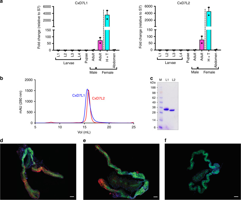
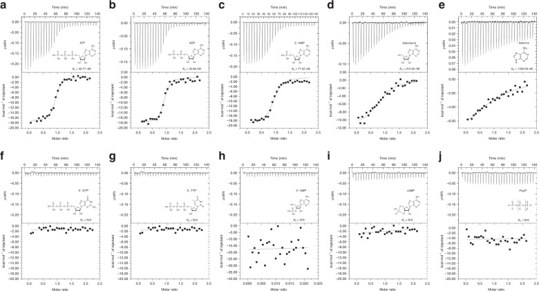
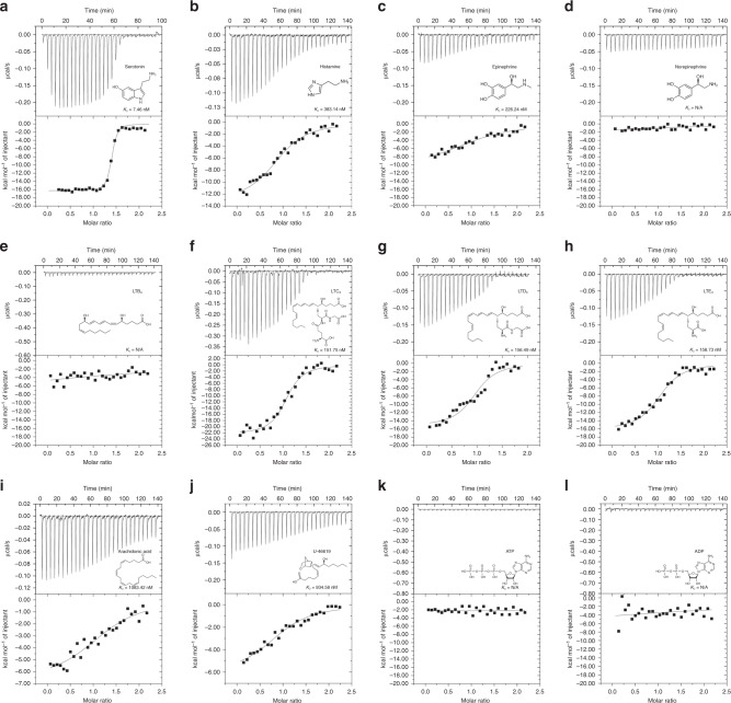
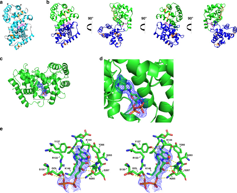
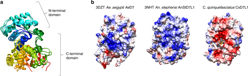
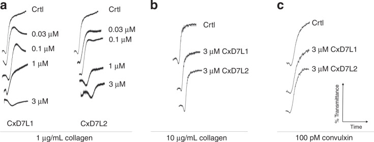
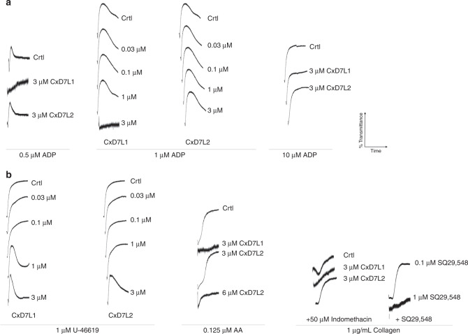
During blood-feeding, mosquito saliva is injected into the skin to facilitate blood meal acquisition. D7 proteins are among the most abundant components of the mosquito saliva. Here we report the ligand binding specificity and physiological relevance of two D7 long proteins from Culex quinquefasciatus mosquito, the vector of filaria parasites or West Nile viruses. CxD7L2 binds biogenic amines and eicosanoids. CxD7L1 exhibits high affinity for ADP and ATP, a binding capacity not reported in any D7. We solve the crystal structure of CxD7L1 in complex with ADP to 1.97 Å resolution. The binding pocket lies between the two protein domains, whereas all known D7s bind ligands either within the N- or the C-terminal domains. We demonstrate that these proteins inhibit hemostasis in ex vivo and in vivo experiments. Our results suggest that the ADP-binding function acquired by CxD7L1 evolved to enhance blood-feeding in mammals, where ADP plays a key role in platelet aggregation.


Conflict of interest statement

The authors declare no competing interests.

Figures

Fig. 1
Fig. 1. Characterization of C. quinquefasciatus salivary long D7 proteins.
a Gene expression analysis of CxD7L1 and CxD7L2 transcripts in different stages of C. quinquefasciatus mosquitoes. Relative abundance was expressed as the fold change using the 40S ribosomal protein S7 as the housekeeping gene. Larvae stage 1 (L1), larvae stage 2 (L2), larvae stage 3 (L3), larvae stage 4 (L4), pupae, male adult (reference sample), female adult, heads and thoraxes (H+T), and abdomens from female adult mosquitoes were analyzed separately. Two biological replicates and two technical duplicates were analyzed. Bars indicate the standard error of the means. b Purification of CxD7L1 (blue line) and CxD7L2 (red line) by size exclusion chromatography using Superdex 200 Increase 10/300 GL column. c Coomassie-stained NuPAGE Novex 4–12% Bis–Tris gel electrophoresis (n = 1) of recombinant proteins CxD7L1 and CxD7L2 (1.5 µg). SeeBlue Plus2 Pre-stained was used as the protein standard (M). d, e Immunolocalization of CxD7L1 and CxD7L2 proteins in the salivary glands of C. quinquefasciatus. Salivary glands were incubated with rabbit IgG anti-CxD7L1 (d), anti-CxD7L2 (e), and further stained with anti-rabbit IgG Alexa Fluor 594 antibody shown in red. Proteins of interest were localized in the medial and distal regions of the lateral lobes of C. quinquefasciatus salivary glands. As a control, salivary glands were incubated with anti-rabbit IgG AF594 alone (f). Nucleic acids were stained by DAPI (blue) and the actin structure of salivary glands was stained using Phalloidin Alexa 488 (green). Four independent experiments were performed with 1–2 glands imaged per experimental group. Scale bar = 50 µm. Source data are provided as a Source Data file.
Fig. 2
Fig. 2. Binding of nucleosides and related molecules to CxD7L1 by ITC.
Binding experiments were performed on a VP-ITC microcalorimeter. Assays were performed at 30 °C. The upper curve in each panel shows the measured heat for each injection, while the lower graph shows the enthalpies for each injection and the fit to a single-site binding model for calculation of thermodynamic parameters. Titration curves are representative of two measurements. Panels (ae) show adenine nucleosides or nucleotides that bind CxD7L1: adenosine 5-triphosphate (a), adenosine 5-diphosphate (b), adenosine 5-monophosphate (c), adenosine (d), and adenine (e). In panels (fj), other purine and pyrimidine nucleotides and related substances showed no binding to CxD7L1: guanosine 5-triphosphate (f), thymidine 5-triphosphate (g), adenosine 3-monophosphate (h), cyclic adenosine monophosphate (i), and polyphosphate (j). The insets show the names and chemical formulas for these compounds.
Fig. 3
Fig. 3. Binding of biogenic amines and eicosanoids to CxD7L2 by ITC.
Binding experiments were performed on a VP-ITC microcalorimeter. The upper curve in each panel shows the measured heat for each injection, while the lower graph shows the enthalpies for each injection and the fit to a single-site binding model for calculation of thermodynamic parameters. Titration curves are representative of two measurements. Panels: serotonin (a), histamine (b), epinephrine (c), norepinephrine (d), LTE4 (e), LTC4 (f), LTD4 (g), LTE4 (h), arachidonic acid (i), TXA2 analog U-46619 (j), ATP (k), and ADP (l). The insets show the names and chemical formulas for these compounds.
Fig. 4
Fig. 4. Structure of CxD7L1 in complex with ADP.
a Ribbon representation of CxD7L1-ADP structure. The 17 α-helices are labeled A–Q. b Several views of CxD7L1 differing by rotations of 90° around the y-axis. N-terminal and C-terminal are colored in blue and green, respectively. ADP is shown as a stick model in magenta and disulfide bonds in orange. c Electron density map covering ADP. CxD7L1 protein is colored in green. d Inset from (c) is shown. Amino acid residues of CxD7L1 involved in ADP binding are colored in green (e). Stereo view of the binding pocket of the CxD7L1-ADP complex showing the 2Fo − Fc electron density contoured at 1σ covering the ligand. All residues within a 3.6 Å distance from the ADP are shown. Hydrogen bonds are colored in yellow. GOL-402 is a glycerol molecule that is in the area of the phosphate group. R271 was not included in the figure as it is not within 3.6 Å and it binds N265, but it does not bind ADP directly.
Fig. 5
Fig. 5. Multiple sequence superposition and electrostatic potential of Culex D7 proteins.
a Superposition of CxD7L1, Ae. aegypti AeD7 (PDB ID: 3DZT) and An. stephensi AnStD7L1 (PDB ID: 3NHT) shows a similar overall helix structure. Rainbow coloring pattern shows the N-terminal in blue and the C-terminal in red. b Electrostatic potential of 3DZT, 3NHT, and CxD7L1 generated by Coulombic Surface Coloring (Chimera software) with blue being positive and red being negative. ADP is represented as a stick model.
Fig. 6
Fig. 6. Effect of CxD7 proteins on platelet aggregation induced by collagen or convulxin.
Prior to the addition of the agonist, platelet-rich human plasma was incubated for 1 min with either PBS (Crtl) or with the recombinant proteins at the concentrations shown. Aggregometer traces were measured at 37 °C from stirred platelets suspensions on a Chrono-Log platelet aggregometer model 700 for 6 min. An increase of light transmittance over time indicates platelet aggregation. a CxD7L1 and CxD7L2 concentration-dependent inhibition of platelet aggregation induced by low doses of collagen (1 µg/mL). CxD7L1 and CxD7L2 failed to inhibit platelet aggregation induced by b high doses of collagen (10 µg/mL) and c GPVI agonist convulxin (100 pM). Graphs are representative of two measurements. Source data are provided as a Source Data file.
Fig. 7
Fig. 7. Effect of CxD7 proteins on platelet aggregation induced by secondary mediators.
Prior to the addition of the agonist, platelet-rich human plasma was incubated for 1 min either with PBS (Crtl) or with the recombinant proteins, or SQ29,548 at the concentrations shown. Aggregometer traces were measured at 37 °C from stirred platelets suspensions on a Chrono-Log platelet aggregometer model 700 for 6 min. An increase of light transmittance over time indicates platelet aggregation. a Platelet aggregation traces using different concentrations of ADP (0.5, 1, and 10 µM) as aggregation agonist. b Platelet aggregation traces using 1 µM U-46619, 0.125 µM arachidonic acid (AA), or low collagen concentration (1 µg/mL). Graphs are representative of two measurements. Source data are provided as a Source Data file.

References

    1. Lai CH, Tung KC, Ooi HK, Wang JS. Competence of Aedes albopictus and Culex quinquefasciatus as vector of Dirofilaria immitis after blood meal with different microfilarial density. Vet. Parasitol. 2000;90:231–237. - PubMed
    1. Simonsen PE, Mwakitalu ME. Urban lymphatic filariasis. Parasitol. Res. 2013;112:35–44. - PMC - PubMed
    1. Cornet S, Nicot A, Rivero A, Gandon S. Evolution of plastic transmission strategies in Avian Malaria. PLoS Pathog. 2014;10:e1004308. - PMC - PubMed
    1. Farajollahi A, Fonseca DM, Kramer LD, Kilpatrick AM. “Bird biting” mosquitoes and human disease: a review of the role of Culex pipiens complex mosquitoes in epidemiology. Infect. Genet. Evol. 2011;11:1577–1585. - PMC - PubMed
    1. Turell MJ. Members of the Culex pipiens complex as vectors of viruses. J. Am. Mosq. Control Assoc. 2012;28:123–126. - PubMed

Publication types

MeSH terms