Review
. 2020 Jul;21(7):366-383.
doi: 10.1038/s41583-020-0314-2. Epub 2020 Jun 9.

Transplanting neural progenitor cells to restore connectivity after spinal cord injury

Affiliations
Review

Transplanting neural progenitor cells to restore connectivity after spinal cord injury

Itzhak Fischer et al. Nat Rev Neurosci. 2020 Jul.

Abstract

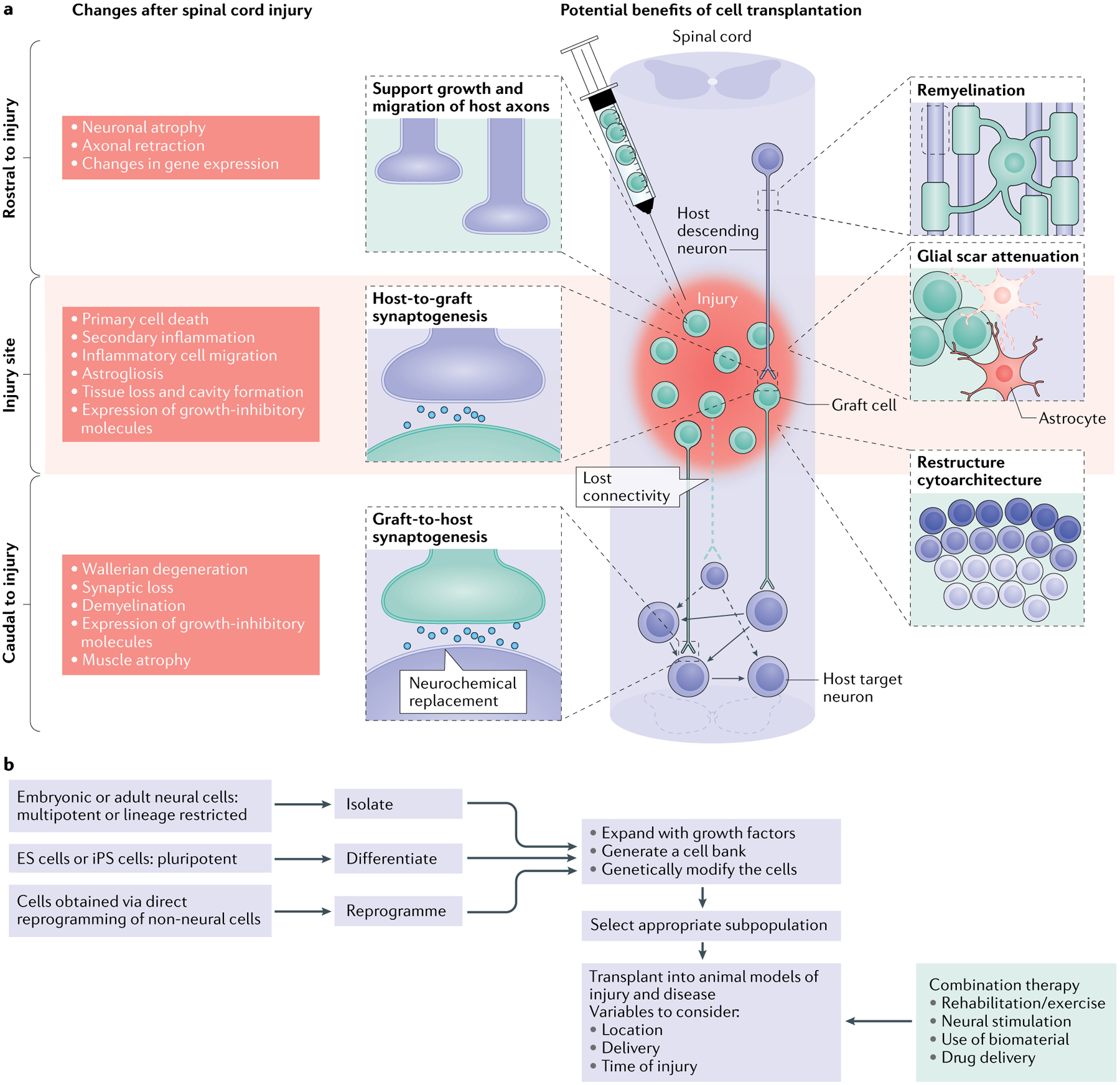
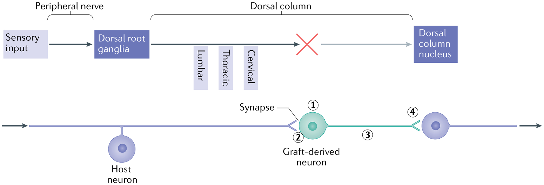
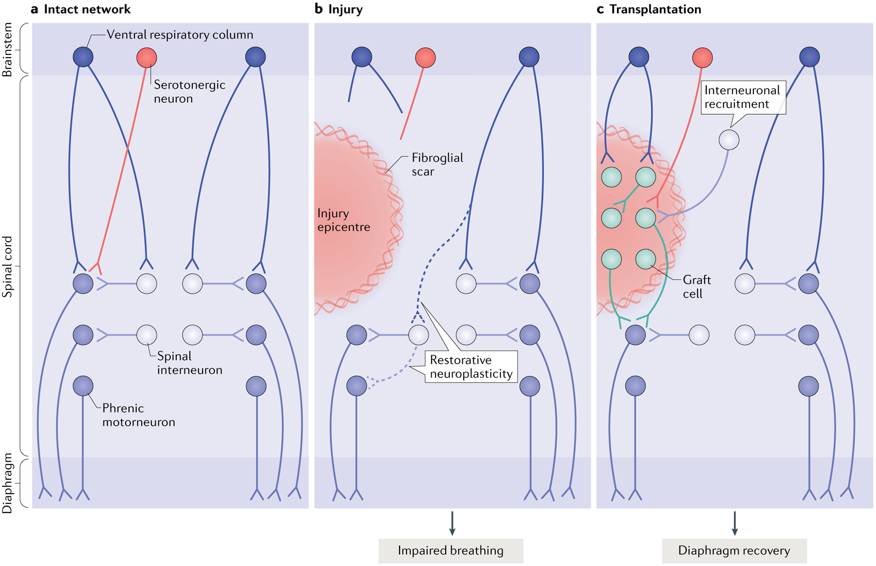
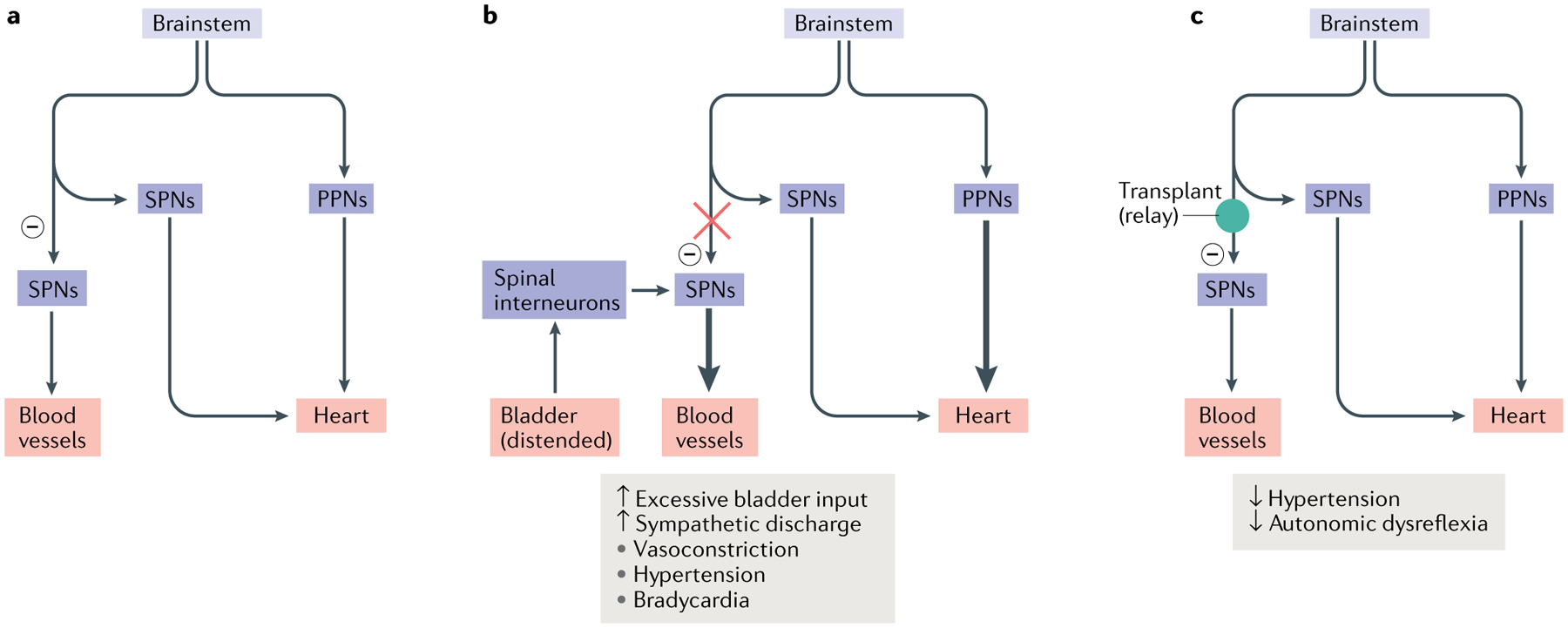
Spinal cord injury remains a scientific and therapeutic challenge with great cost to individuals and society. The goal of research in this field is to find a means of restoring lost function. Recently we have seen considerable progress in understanding the injury process and the capacity of CNS neurons to regenerate, as well as innovations in stem cell biology. This presents an opportunity to develop effective transplantation strategies to provide new neural cells to promote the formation of new neuronal networks and functional connectivity. Past and ongoing clinical studies have demonstrated the safety of cell therapy, and preclinical research has used models of spinal cord injury to better elucidate the underlying mechanisms through which donor cells interact with the host and thus increase long-term efficacy. While a variety of cell therapies have been explored, we focus here on the use of neural progenitor cells obtained or derived from different sources to promote connectivity in sensory, motor and autonomic systems.


Conflict of interest statement

Competing interests

The authors declare no competing interests.

Figures

Fig. 1 |
Fig. 1 |. Spinal cord injury: pathophysiological events and potential therapeutic targets.
a | The left side of the schematic illustrates the complex changes that occur after spinal cord injury (SCI), which differ temporally and spatially. In a descending tract, such as that illustrated here, these changes include events rostral to the injury (including axon degeneration and changes in gene expression), at the level of the injury (encompassing acute tissue damage and cell death as well as chronic secondary injury and inflammation) and caudal to the injury (including both neural events such as demyelination and non-neural events such as muscle atrophy). Similar changes occur in ascending tracts; however, in this case the location of the events in relation to the injury will be reversed. The injured spinal cord schematic illustrates potential therapeutic targets for cell transplantation, including remyelination, support of host axon growth, glial scar attenuation, synaptogenesis and the restructuring of spinal cord cytoarchitecture. b | The flow chart depicts the decisions that must be made when a cell transplantation strategy for SCI is being developed and the processing steps involved. It shows choices of cells for transplantation in SCI, the process of their preparation, modification and selection and the parameters of their delivery alone and as part of a combination therapy. For cell choices, a wide range of neural progenitor cells can be obtained from embryonic and adult tissue, from pluripotent cells (embryonic stem cells (ES cells) and induced pluripotent stem cells (iPS cells)) and from direct reprogramming of non-neural cells. These cells can be expanded with growth factors to generate cell banks and/or can be genetically modified (to overexpress growth factors, for example). It is then possible to select a subpopulation of the resulting neurons for transplantation. The transplantation process needs to consider variables such as the location of the transplant (that is, transplantation directly into the injury site, intrathecally or systemically), the delivery method (that is, injection as a cell suspension or as part of a hydrogel scaffold) and the timing (for example, subacute transplantation versus transplantation after a 2-week delay after injury). A number of different types of combination therapy can also be initiated at different times and act synergistically with the transplant. In particular, attention should be paid to rehabilitation strategies, various neural stimulation modalities, the use of biomaterials and drug delivery.
Fig. 2 |
Fig. 2 |. Forming a relay using neural progenitor cells.
The schematic illustrates the elements required to use neural progenitor cells (NPCs) to form a relay following spinal cord injury, using the example of the sensory system in the presence of a cervical lesion of the dorsal column and transplants of neuronal-restricted precursors (NRPs) and/or glial-restricted precursors (GRPs). The upper part illustrates the elements of the ascending sensory pathway, disrupted by a cervical injury that interrupts the connectivity of sensory axons (dorsal column) with the dorsal column nucleus (DCN) in the brainstem. The lower part shows the steps required to restore the connectively using NPC transplants that form a relay. First, the transplant must survive and generate neurons with the appropriate phenotype (excitatory, for example) (1). The use of a mixture of NRPs and GRPs has been found to be effective, as the GRP-derived astrocytes generate a permissive environment for survival and differentiation of neurons. Second, the host axons must grow into the graft and form synaptic connections (2). It appears that the presence of astrocytes in the graft attracts the sensory neurons, but other strategies include the induction of the growth potential of host neurons through the repression of genes such as PTEN and SOCS3 (REF.). Finally, the axons of transplanted neurons must undergo directional extension to the target (along a neurotrophic gradient to the DCN) (3) and form synaptic connections (4). To verify the formation of a functional relay, analysis needs to be performed at different levels. Structural analysis includes the tracing of axon growth from the host and the transplant and obtaining evidence of synaptic structure by electron microscopy. Physiological analysis may involve the stimulation of axons followed by assessment of the expression of FOS in downstream neurons as well as measures of signal transmission through the transplant. Functional analysis will include behaviour tests indicative of restored connectivity of the specific tracts.
Fig. 3 |
Fig. 3 |. Restoring connectivity in the respiratory system.
The diagram depicts the intact (part a), injured (part b) and transplant-treated (part c) spinal phrenic motor circuit within the cervical spinal cord. Respiration is driven by brainstem neurons in the ventral respiratory column that directly — or indirectly via spinal interneurons — innervate the phrenic motor neuron pool (distributed from cervical level C3 to cervical level C6). Phrenic motor neuron activity is also modulated by serotonergic pathways and populations of spinal interneurons as breathing conditions change. Phrenic motor neurons on each side of the spinal cord innervate half of the diaphragm on each side of the body via phrenic nerves. Injury (part b) can compromise descending projections, as well as phrenic spinal interneurons and motor neurons. Spared spinal neurons caudal to the injury are therefore denervated. While this is devastating, some limited recovery of diaphragm activity can occur ipsilateral to the injury via restorative neuroplasticity (dashed lines), and spared monosynaptic and polysynaptic pathways from the contralateral spinal cord (via brainstem and spinal interneurons, respectively) can facilitate plasticity in these lateralized spinal injuries. However, the extent of recovery is minimal and deficits persist. A number of cell therapies have been used to promote repair and plasticity within injured respiratory pathways. Neural progenitor cell transplants are perhaps the most often used, as they can modify glial scarring at the lesion border and provide the building blocks for tissue repair. Transplantation of neural progenitor cells into the injured phrenic network in animal models (typically directly into the lesion site as shown in part c) has resulted in extensive synaptic integration between donor neurons themselves, between host spinal and brainstem neurons and donor neurons and between donor and spinal phrenic neurons. This synaptic integration also coincides with enhanced plasticity of existing and newly formed pathways, and improved respiratory activity,,,. Without any other intervention, transplantation of cells alone is likely to lead to the formation of a vast range of new connections, which are likely to differ between treated recipients,, and to the recruitment of novel interneuron populations to establish novel neural networks. Research is under way to develop strategies that control this integration and connectivity. Other models of injury affecting the phrenic network and additional mechanisms of recovery are discussed elsewhere.
Fig. 4 |
Fig. 4 |. Restoring connectivity in autonomic systems.
The majority of the vasculature is controlled by sympathetic activity, while the heart is regulated by both the sympathetic system and the parasympathetic system. Sympathetic preganglionic neurons (SPNs) in the spinal cord project to the periphery and synapse onto sympathetic postganglionic neurons. The latter extend axon terminals into the blood vessel and heart. a | In normal conditions, sympathetic excitation induces vasoconstriction and thus increases blood pressure. Subsequently, baroreceptor-mediated parasympathetic excitation decreases the heart rate. In addition, supraspinal vasomotor pathways provide inhibitory regulation (indicated by a minus sign) to suppress the sympathetic activity to blood vessels, leading to recovery of normal blood pressure. b | After spinal cord injury, spinal SPNs lose this descending inhibitory modulation. When excessive sensory or visceral stimulation below the level of injury (for example, bladder distension) activates SPNs via interneurons, the massive discharge of SPNs causes vasoconstriction and increases blood pressure. This causes baroreceptor-mediated bradycardia to occur. However, the absence of supraspinal inhibitory signals to caudal SPNs means that blood pressure remains high. The resulting simultaneous hypertension and bradycardia is known as autonomic dysreflexia. c | Transplantation of early-stage neurons into the lesion of the spinal cord reconstitutes supraspinal vasomotor pathways. Grafted cells relay supraspinal inhibitory signals across the lesion to target neurons in the caudal portion of the spinal cord, which can restore sympathetic regulation of cardiovascular function after spinal cord injury. PPNs, parasympathetic preganglionic neurons.

Comment in

  • What the Editors are reading - Basic science.
    Wu HY, Willacy O, Fossum M. Wu HY, et al. J Pediatr Urol. 2021 Apr;17(2):271-272. doi: 10.1016/j.jpurol.2020.12.010. Epub 2020 Dec 10. J Pediatr Urol. 2021. PMID: 33358556 No abstract available.

References

    1. Ahuja CS et al.Traumatic spinal cord injury. Nat. Rev. Dis. Prim 3, 17018 (2017). - PubMed
    1. Mautes AE, Weinzierl MR, Donovan F & Noble LJ Vascular events after spinal cord injury: contribution to secondary pathogenesis. Phys. Ther 80, 673–687 (2000). - PubMed
    1. Beattie MS Inflammation and apoptosis: linked therapeutic targets in spinal cord injury. Trends Mol. Med 10, 580–583 (2004). - PubMed
    1. Donnelly DJ & Popovich PG Inflammation and its role in neuroprotection, axonal regeneration and functional recovery after spinal cord injury. Exp. Neurol 209, 378–388 (2008). - PMC - PubMed
    1. Jia Z et al.Oxidative stress in spinal cord injury and antioxidant-based intervention. Spinal Cord 50, 264–274 (2012). - PubMed

Publication types