

doi: 10.12688/f1000research.21294.1.
eCollection 2019.
Adapting the 3D-printed Openflexure microscope enables computational super-resolution imaging
Affiliations
- PMID: 32518624
- PMCID: PMC7255852
- DOI: 10.12688/f1000research.21294.1
Item in Clipboard
Adapting the 3D-printed Openflexure microscope enables computational super-resolution imaging
F1000Res.
.
Abstract
We report on a 3D printed microscope, based on a design by the Openflexure project, that uses low cost components to perform fluorescence imaging. The system is sufficiently sensitive and mechanically stable to allow the use of the Super Resolution Radial Fluctuations algorithm to obtain images with resolution better than the diffraction limit. Due to the low-cost components, the entire system can be built for approximately $1200.
Keywords: 3D Printing; Light Microscopy; Open Science; Super-resolution.
Copyright: © 2019 Grant SD et al.
Conflict of interest statement
No competing interests were disclosed.
Figures
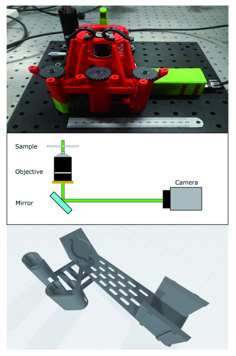
Top - image of microscope as constructed. It is mounted on an aluminium breadboard for portability and stability. The ruler is 15cm and shown for scale. The colours of individual parts are arbitrary. Middle - beam path showing key design parameters. Illumination is from a laser mounted above the sample and the camera is mounted at the primary imaging plane of the microscope objective with an emission filter directly attached. The turning mirror allows a more compact design. Bottom: integrated mirror and camera mount to allow imaging with high sensitivity camera. We used the slots in the base of this part to pass mounting screws to the breadboard for additional stability.
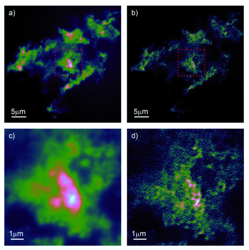
a) Drift corrected and summed stack of 200 individual frames.
b) Superresolution radial fluctuations (SRRF)-processed image of same stack of images.
c),
d) zoom on equivalent region marked by red dashed box in
a),
b) showing increased resolution enabled by SRRF.

The nanodiamonds were suspended in the growth medium before uptake by the macrophages.
a),
b) large field of view encompassing multiple macrophages in both wide-field fluorescence and super-resolution radial fluctuations (SRRF) imaging modes.
c),
d) detail view of single macrophage in both widefield and SRRF modes.
References
MeSH terms
LinkOut - more resources
Full Text Sources