

Correlation Between Chest CT Findings and Clinical Features of 211 COVID-19 Suspected Patients in Wuhan, China
- PMID: 32518804
- PMCID: PMC7239186
- DOI: 10.1093/ofid/ofaa171
Correlation Between Chest CT Findings and Clinical Features of 211 COVID-19 Suspected Patients in Wuhan, China
Abstract
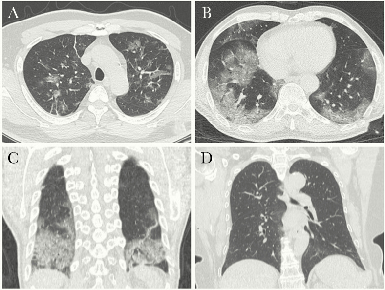
Background: Chest computed tomography (CT) has been widely used to assess pulmonary involvement in COVID-19. We aimed to investigate the correlation between chest CT and clinical features in COVID-19 suspected patients with or without fever.
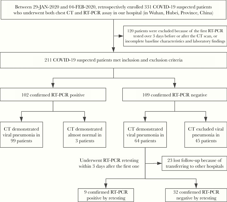
Methods: We retrospectively enrolled 211 COVID-19 suspected patients who underwent both chest CT and reverse transcription polymerase chain reaction in Wuhan, China. The performance of CT in patients with relevant onset of symptoms, with fever (n = 141) and without fever (n = 70), was assessed respectively.
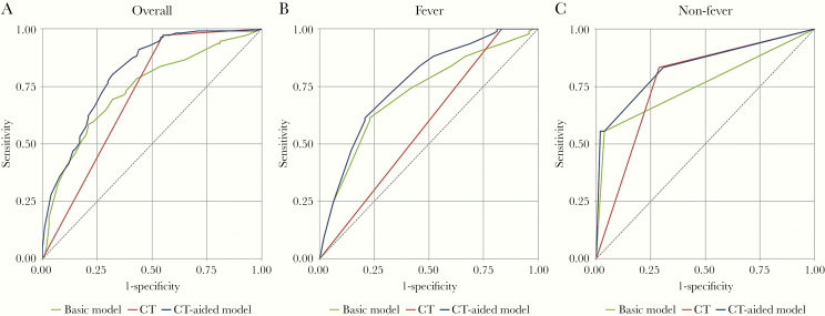
Results: The sensitivity of CT for COVID-19 was 97.3%, with area under the curve (AUC) of 0.71 (95% confidence interval [CI], 0.66-0.76). There were 141 suspected patients with fever and 70 without fever. In the fever group, 4 variables were screened to establish the basic model: age, monocyte, red blood cell, and hypertension. The AUC of the basic model was 0.72 (95% CI, 0.63-0.81), while the AUC of the CT-aided model was 0.77 (95% CI, 0.68-0.85), a significant difference (P < .05). In the nonfever group, only dry cough was screened out to establish the basic model. The AUC was 0.76 (95% CI, 0.64-0.88), which was not significantly different than the CT-aided model (P = .08).
Conclusions: Chest CT has a high sensitivity in patients with COVID-19, and it can improve diagnostic accuracy for COVID-19 suspected patients with fever during the initial screen, whereas its value for nonfever patients remains questionable.
Keywords: COVID-19; polymerase chain reaction; reverse transcriptase ROC curve; tomography; x-ray computed.
© The Author(s) 2020. Published by Oxford University Press on behalf of Infectious Diseases Society of America.
Figures
Similar articles
-
Thoracic imaging tests for the diagnosis of COVID-19.Cochrane Database Syst Rev. 2020 Sep 30;9:CD013639. doi: 10.1002/14651858.CD013639.pub2. Cochrane Database Syst Rev. 2020. Update in: Cochrane Database Syst Rev. 2020 Nov 26;11:CD013639. doi: 10.1002/14651858.CD013639.pub3. PMID: 32997361 Updated.
-
Thoracic imaging tests for the diagnosis of COVID-19.Cochrane Database Syst Rev. 2020 Nov 26;11:CD013639. doi: 10.1002/14651858.CD013639.pub3. Cochrane Database Syst Rev. 2020. Update in: Cochrane Database Syst Rev. 2021 Mar 16;3:CD013639. doi: 10.1002/14651858.CD013639.pub4. PMID: 33242342 Updated.
-
Diagnostic performance between CT and initial real-time RT-PCR for clinically suspected 2019 coronavirus disease (COVID-19) patients outside Wuhan, China.Respir Med. 2020 Jul;168:105980. doi: 10.1016/j.rmed.2020.105980. Epub 2020 Apr 21. Respir Med. 2020. PMID: 32364959 Free PMC article.
-
A comparison of clinical, laboratory and chest CT findings of laboratory-confirmed and clinically diagnosed COVID-19 patients at first admission.Diagn Interv Radiol. 2021 May;27(3):336-343. doi: 10.5152/dir.2020.20270. Diagn Interv Radiol. 2021. PMID: 32876570 Free PMC article.
-
Correlation between Chest Computed Tomography and Lung Ultrasonography in Patients with Coronavirus Disease 2019 (COVID-19).Ultrasound Med Biol. 2020 Nov;46(11):2918-2926. doi: 10.1016/j.ultrasmedbio.2020.07.003. Epub 2020 Jul 13. Ultrasound Med Biol. 2020. PMID: 32771222 Free PMC article.
Cited by
-
Analytic and Clinical Performance of Major Commercial Severe Acute Respiratory Syndrome Coronavirus 2 Molecular Assays in the United States.Clin Lab Med. 2022 Jun;42(2):129-145. doi: 10.1016/j.cll.2022.02.001. Epub 2022 Feb 21. Clin Lab Med. 2022. PMID: 35636818 Free PMC article. Review.
-
[Korean Clinical Imaging Guidelines for Justification of Diagnostic Imaging Study for COVID-19].Taehan Yongsang Uihakhoe Chi. 2022 Mar;83(2):265-283. doi: 10.3348/jksr.2021.0117. Epub 2022 Jan 6. Taehan Yongsang Uihakhoe Chi. 2022. PMID: 36237918 Free PMC article. Review. Korean.
-
Thoracic imaging tests for the diagnosis of COVID-19.Cochrane Database Syst Rev. 2022 May 16;5(5):CD013639. doi: 10.1002/14651858.CD013639.pub5. Cochrane Database Syst Rev. 2022. PMID: 35575286 Free PMC article.
-
An Umbrella Review With Meta-Analysis of Chest Computed Tomography for Diagnosis of COVID-19: Considerations for Trauma Patient Management.Front Med (Lausanne). 2022 Jul 26;9:900721. doi: 10.3389/fmed.2022.900721. eCollection 2022. Front Med (Lausanne). 2022. PMID: 35957847 Free PMC article.
-
Accurately Differentiating Between Patients With COVID-19, Patients With Other Viral Infections, and Healthy Individuals: Multimodal Late Fusion Learning Approach.J Med Internet Res. 2021 Jan 6;23(1):e25535. doi: 10.2196/25535. J Med Internet Res. 2021. PMID: 33404516 Free PMC article.
References
-
- World Health Organization. Clinical management of severe acute respiratory infection (SARI) when COVID-19 disease is suspected. Available at: https://www.who.int/publications-detail/clinical-management-of-severe-ac.... Accessed 11 March 2020.
