. 2020 May 13:17:1202-1214.
doi: 10.1016/j.omtm.2020.05.008. eCollection 2020 Jun 12.

Metformin, an AMPK Activator, Inhibits Activation of FLSs but Promotes HAPLN1 Secretion

Affiliations

Metformin, an AMPK Activator, Inhibits Activation of FLSs but Promotes HAPLN1 Secretion

Yong Chen et al. Mol Ther Methods Clin Dev. .

Abstract

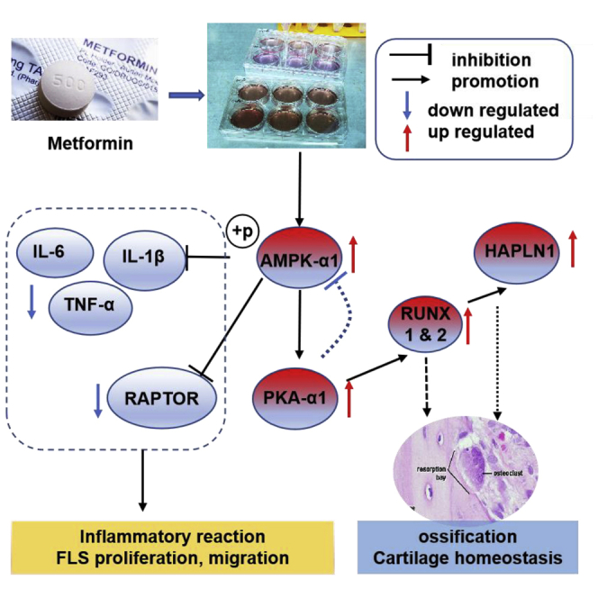
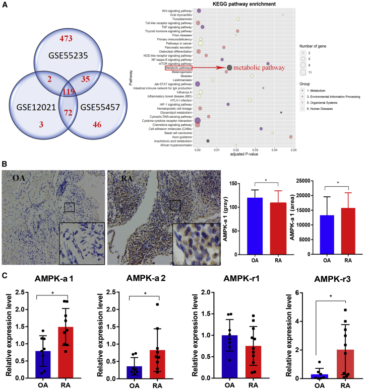
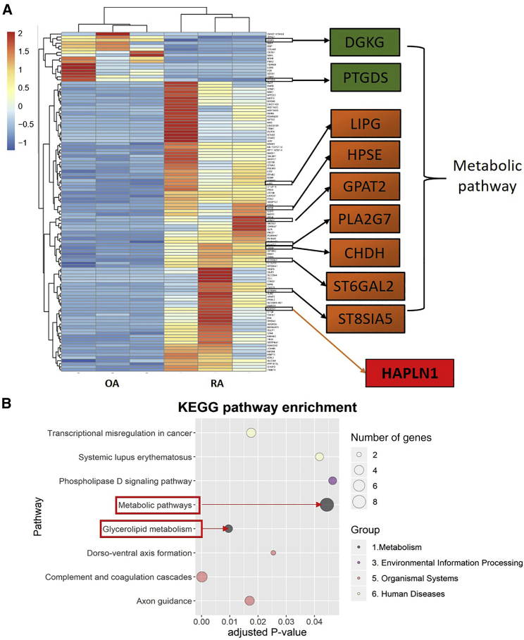
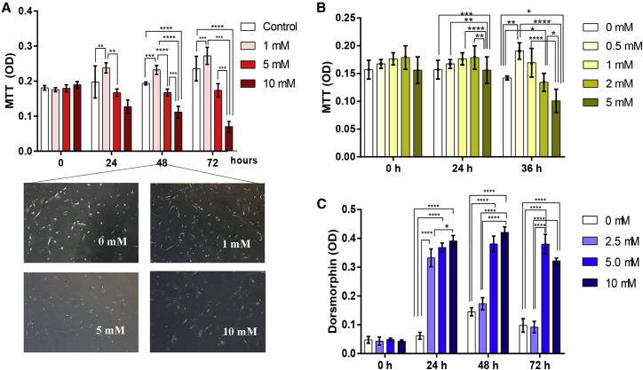
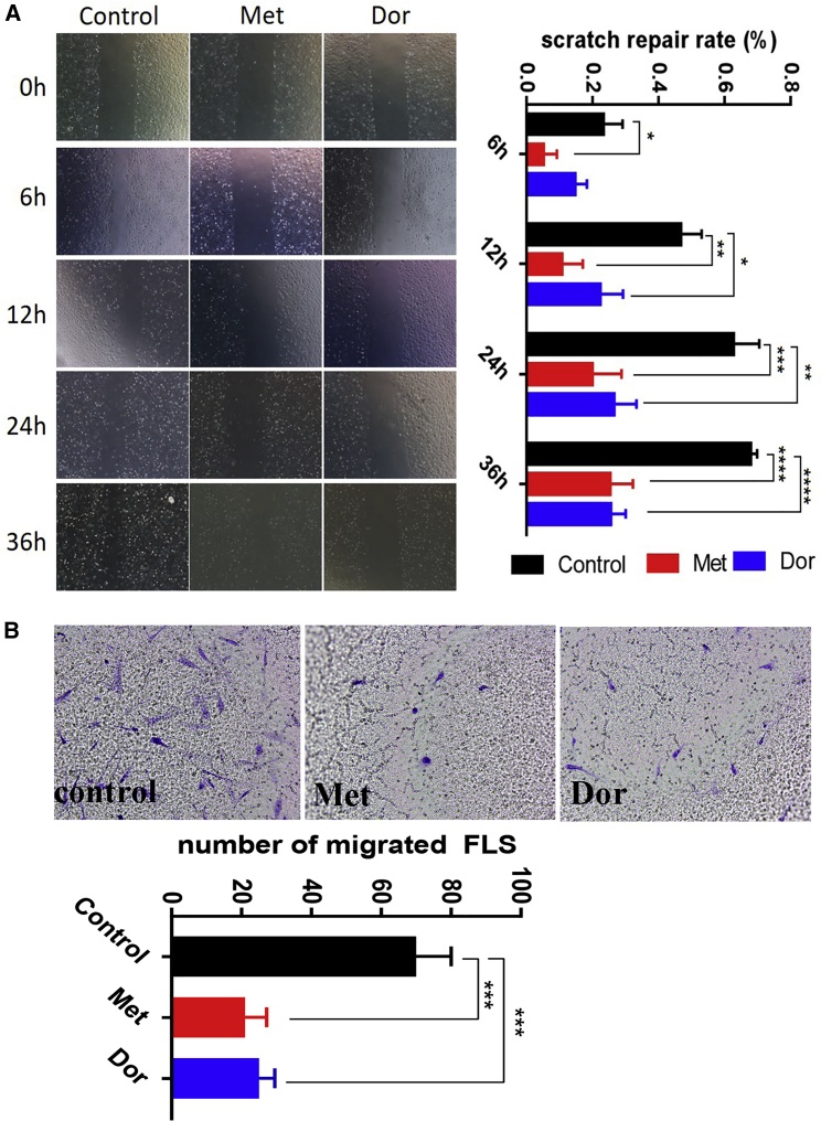
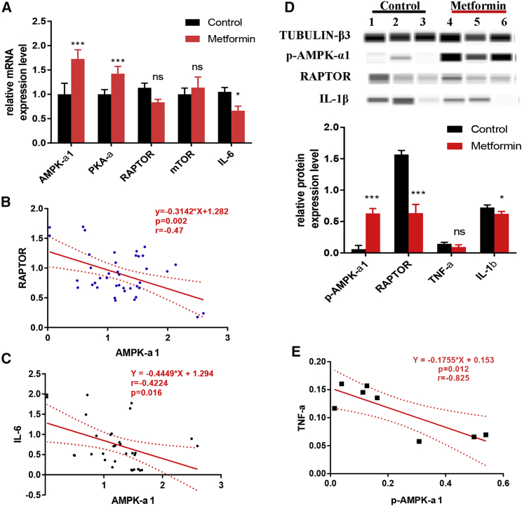
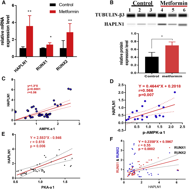
AMP-activated protein kinase (AMPK) is essential for maintaining energy balance and has a crucial role in various inflammatory pathways. In this study, AMPK levels positively correlated with many inflammatory indexes in rheumatoid arthritis (RA) patients, especially in the affected synovium. In RA sera, a positive correlation between phosphorylated (p-)AMPK-α1 levels and DAS28 (disease activity score 28) activity (r = 0.270, p < 0.0001) was found. Similarly, a positive correlation was observed between AMPK-α1 and tumor necrosis factor α (TNF-α) levels (r = 0.460, p = 0.0002). Differentially expressed genes between osteoarthritis (OA) and RA synovium from NCBI GEO profiles and our RNA sequencing data suggested activation of metabolic pathways specific to RA-fibroblast-like synoviocytes (FLSs). AMPK-α1 was highly expressed in the synovium of RA but not OA patients. An AMPK activator, metformin, inhibited FLS proliferation at higher but not lower concentrations, whereas the inhibitor dorsomorphin promoted the proliferation of RA-FLSs. Interestingly, both metformin and dorsomorphin inhibited the migration of RA-FLSs. After metformin treatment, expression of interleukin 6 (IL-6), TNF-α, and IL-1β were significantly downregulated in RA-FLSs; however, increased expression of p-AMPK-α1, protein kinase A (PKA)-α1, and HAPLN1 (hyaluronan and proteoglycan link protein 1) was observed. Increased levels of HAPLN1 in RA-FLSs by an AMPK activator could potentially be beneficial in protecting the joints. Hence, our results demonstrate the potential of an AMPK activator as a therapeutic for RA.

Keywords: AMP-activated protein kinase; disease activity; fibroblast-like synoviocyte; hyaluronan and proteoglycan link protein 1; metabolism; metformin; rheumatoid arthritis; therapeutic target.


Figures

None
Graphical abstract
Figure 1
Figure 1
Expression of Serum AMPK-α1 and p-AMPK-α1 Levels in RA and OA Patients (A) AMPK-α1 levels between RA (n = 61) and OA (n = 20) patients having moderate to high disease activity were shown, but no statistical significance between groups was observed. (B) Serum p-AMPK-α1 level was higher in OA compared to RA patients having lower disease activity. Log-transformed p-AMPK-α1 values were significantly higher in OA than RA patients having lower disease activity. (C) In RA, patients with higher disease activity had significantly higher levels of log-transformed p-AMPK-α1 levels compared to patients with lower disease activity. (D and E) Log p-AMPK-α1 levels significantly correlated with DAS28 (D) and CRP (E) levels. (F) Correlation with ESR levels was not statistically significant. (G and H) Increased expression of IL-17 (G) and TNF-α (H) was observed in RA than OA patients. (I) Levels of AMPK-α1 moderately correlated with TNF-α levels. ∗p < 0.05, ∗∗p < 0.01, ∗∗∗p < 0.001, ∗∗∗∗p < 0.0001.
Figure 2
Figure 2
Changes in the Metabolic Pathway in RA Compared to OA Synovium (A) In total, 119 DEGs between RA and OA patients’ synovium intersected in all the three datasets (NCBI GEO profiles; GEO: GSE12021, GSE55235, and GSE55457). Among them, changes in the metabolic pathway-related nine genes were more obvious. (B) IHC staining for p-AMPK-α1 was more intense in RA (n = 20) than in OA (n = 17) synovium. (C) Relative expression levels of AMPK-α1, AMPK-α2, and AMPK-γ3 genes were higher in RA (n = 10) than in OA (n = 9) synovium. ∗p < 0.05.
Figure 3
Figure 3
DEGs in FLSs Identified by mRNA High-Throughput Sequencing Method (A) In total, 111 DEGs were identified in RA-FLSs compared to OA samples (n = 3/group). Expression of 95 genes was upregulated compared to the downregulation in the expression of 16 genes. Of all of the nine DEGs related to metabolism, expression levels of DGKG and PTGDS genes were downregulated, while the expression of LIPG, HPSE, GPAT2, PLA2G7, CHDH, ST6GAL2, and ST8SIA5 genes were upregulated in RA-FLSs compared to OA samples. HAPLN1 expression was most obviously upregulated in RA-FLSs. (B) KEGG pathway enrichment analysis demonstrated that the metabolic pathway is one of the most obviously changed pathways.
Figure 4
Figure 4
Effect of AMPK Modulators on RA-FLS Proliferation (A) An MTT assay was used to determine the effects of metformin on RA-FLS viability. Metformin inhibited RA-FLS proliferation at 5 and 10 mM, whereas at 1 mM it has an opposite effect compared to the control group (n = 5). (B) This result was confirmed by using even lower concentration of metformin (n = 5). (C) Dorsomorphin promoted FLS proliferation significantly at 5 and 10 μM compared to the control group after culturing for 36 h (n = 5). A representative example from three independent experiments is shown. ∗p < 0.05, ∗∗p < 0.01, ∗∗∗p < 0.001, ∗∗∗∗p < 0.0001.
Figure 5
Figure 5
AMPK Modulators Inhibited Migration of RA-FLSs (A) A scratch test was performed to evaluate the effects of metformin and dorsomorphin on FLS migration, represented as scratch repair rate (%). Both the AMPK activator (metformin) and inhibitor (dorsomorphin) inhibited FLS migration significantly (n = 3). (B) A transwell assay confirmed the effects of metformin and dorsomorphin on FLS migration after treatment for 24 h (n = 3). A representative example from three independent experiments is shown. ∗p < 0.05, ∗∗p < 0.01, ∗∗∗p < 0.001, ∗∗∗∗p < 0.0001.
Figure 6
Figure 6
Metformin Inhibited Inflammatory Cytokines by Promoting AMPK Expression levels of inflammatory cytokines were measured after culturing RA-FLSs with normal saline or metformin for 36 h (n = 3). (A) Compared to controls, expression levels of AMPK-α1 and PKA-α1 genes were significantly increased while the expression of IL-6 was significantly decreased in metformin-treated RA-FLSs. (B and C) Negative correlation of AMPK-α1 with RAPTOR (B) and IL-6 (C) gene expression. (D) Western blot analysis of IL-1β and TNF-α levels. (E) Negative correlation between p-AMPK-α1 and TNF-α levels. A representative example from three independent experiments is shown. ∗p < 0.05, ∗∗p < 0.01, ∗∗∗p < 0.001, ∗∗∗∗p < 0.0001.
Figure 7
Figure 7
Metformin Promoted HAPLN1 in the PKA-α1/RUNX1/RUNX2 Pathway Relative HAPLN1 expression levels were measured after culturing RA-FLSs with normal saline or metformin for 36 h (n = 3). (A and B) HAPLN1 was increased significantly at both mRNA (A) and protein (B) levels, whereas RUNX1 and RUNX2 were not regulated upon AMPK activation. (C and D) Positive correlation between HAPLN1 and AMPK-α1 at both mRNA (C) and protein (D) levels. (E and F) Positive correlation of HAPLN1 with PKA-α1 (E) and RUNX1 (F) genes. A representative example from three independent experiments is shown. ∗p < 0.05.

References

    1. Calabresi E., Petrelli F., Bonifacio A.F., Puxeddu I., Alunno A. One year in review 2018: pathogenesis of rheumatoid arthritis. Clin. Exp. Rheumatol. 2018;36:175–184. - PubMed
    1. Fazal S.A., Khan M., Nishi S.E., Alam F., Ashraf G.M. A clinical update and global economic burden of rheumatoid arthritis. Endocr. Metab. Immune Disord. Drug Targets. 2018;18:98–109. - PubMed
    1. Bergström B., Lundqvist C., Vasileiadis G.K., Carlsten H., Ekwall O., Ekwall A.H. The rheumatoid arthritis risk gene AIRE is induced by cytokines in fibroblast-like synoviocytes and augments the pro-inflammatory response. Front. Immunol. 2019;10:1384. - PMC - PubMed
    1. Gonzalez-Rey E., Gonzalez M.A., Varela N., O’Valle F., Hernandez-Cortes P., Rico L., Büscher D., Delgado M. Human adipose-derived mesenchymal stem cells reduce inflammatory and T cell responses and induce regulatory T cells in vitro in rheumatoid arthritis. Ann. Rheum. Dis. 2010;69:241–248. - PubMed
    1. Ospelt C., Reedquist K.A., Gay S., Tak P.P. Inflammatory memories: is epigenetics the missing link to persistent stromal cell activation in rheumatoid arthritis? Autoimmun. Rev. 2011;10:519–524. - PubMed