

Acute pulmonary embolism in non-hospitalized COVID-19 patients referred to CTPA by emergency department
- PMID: 32518989
- PMCID: PMC7280685
- DOI: 10.1007/s00330-020-06977-5
Acute pulmonary embolism in non-hospitalized COVID-19 patients referred to CTPA by emergency department
Abstract
Objectives: To evaluate the prevalence of acute pulmonary embolism (APE) in non-hospitalized COVID-19 patients referred to CT pulmonary angiography (CTPA) by the emergency department.
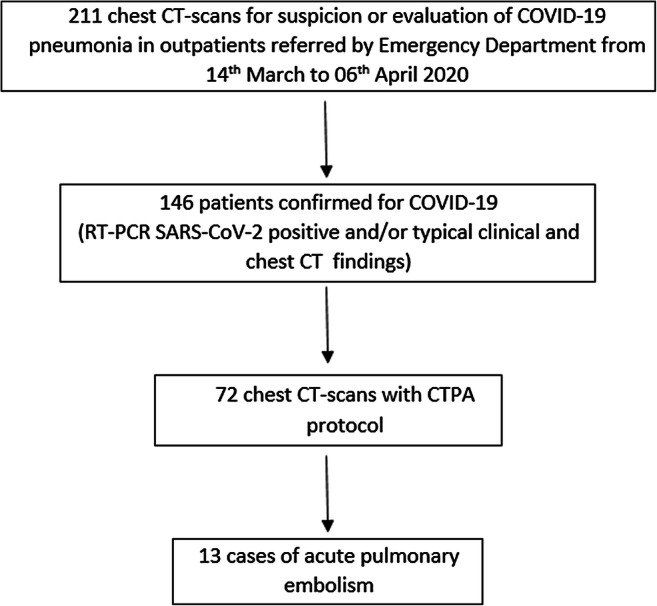
Methods: From March 14 to April 6, 2020, 72 non-hospitalized patients referred by the emergency department to CTPA for COVID-19 pneumonia were retrospectively identified. Relevant clinical and laboratory data and CT scan findings were collected for each patient. CTPA scans were reviewed by two radiologists to determinate the presence or absence of APE. Clinical classification, lung involvement of COVID-19 pneumonia, and CT total severity score were compared between APE group and non-APE group.
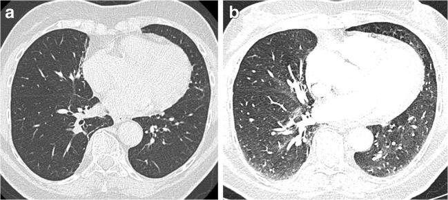
Results: APE was identified in 13 (18%) CTPA scans. The mean age and D-dimer of patients from the APE group were higher in comparison with those from the non-APE group (74.4 vs. 59.6 years, p = 0.008, and 7.29 vs. 3.29 μg/ml, p = 0.011). There was no significant difference between APE and non-APE groups concerning clinical type, COVID-19 pneumonia lung lesions (ground-glass opacity: 85% vs. 97%; consolidation: 69% vs. 68%; crazy paving: 38% vs. 37%; linear reticulation: 69% vs. 78%), CT severity score (6.3 vs. 7.1, p = 0.365), quality of CTPA (1.8 vs. 2.0, p = 0.518), and pleural effusion (38% vs. 19%, p = 0.146).
Conclusions: Non-hospitalized patients with COVID-19 pneumonia referred to CT scan by the emergency departments are at risk of APE. The presence of APE was not limited to severe or critical clinical type of COVID-19 pneumonia.
Key points: • Acute pulmonary embolism was found in 18% of non-hospitalized COVID-19 patients referred by the emergency department to CTPA. Two (15%) patients had main, four (30%) lobar, and seven (55%) segmental acute pulmonary embolism. • Five of 13 (38%) patients with acute pulmonary embolism had a moderate clinical type. • Severity and radiological features of COVID-19 pneumonia showed no significant difference between patients with or without acute pulmonary embolism.
Keywords: CT angiography; Coronavirus; Pneumonia; Pulmonary embolism.
Conflict of interest statement
The authors of this manuscript declare no relationships with any companies, whose products or services may be related to the subject matter of the article.
Figures

References
MeSH terms
LinkOut - more resources
Full Text Sources
Medical
Research Materials
Miscellaneous
