

Chest X-ray in new Coronavirus Disease 2019 (COVID-19) infection: findings and correlation with clinical outcome
- PMID: 32519256
- PMCID: PMC7282464
- DOI: 10.1007/s11547-020-01232-9
Chest X-ray in new Coronavirus Disease 2019 (COVID-19) infection: findings and correlation with clinical outcome
Abstract
Aim: The purpose of this study is to describe the main chest radiological features (CXR) of COVID-19 and correlate them with clinical outcome.
Materials and methods: This is a retrospective study involving patients with clinical-epidemiological suspect of COVID-19 infection, who performed CXRs at the emergency department (ED) of our University Hospital from March 1 to March 31, 2020. All patients performed RT-PCR nasopharyngeal and throat swab, CXR at the ED and clinical-epidemiological data. RT-PCR results were considered the reference standard. The final outcome was expressed as discharged or hospitalized patients into a medicine department or intensive care unit (ICU).
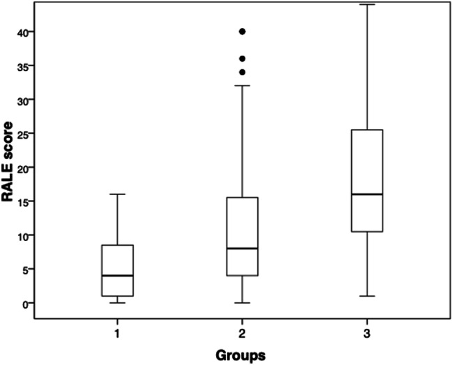
Results: Patients that had a RT-PCR positive for COVID-19 infection were 234 in total: 153 males (65.4%) and 81 females (34.6%), with a mean age of 66.04 years (range 18-97 years). Thirteen CXRs were negative for radiological thoracic involvement (5.6%). The following alterations were more commonly observed: 135 patients with lung consolidations (57.7%), 147 (62.8%) with GGO, 55 (23.5%) with nodules and 156 (66.6%) with reticular-nodular opacities. Patients with consolidations and GGO coexistent in the same radiography were 35.5% of total. Peripheral (57.7%) and lower zone distribution (58.5%) were the most common predominance. Moreover, bilateral involvement (69.2%) was most frequent than unilateral one. Baseline CXR sensitivity in our experience is about 67.1%. The most affected patients were especially males in the age group 60-79 years old (45.95%, of which 71.57% males). RALE score was slightly higher in male than in female patients. ANOVA with Games-Howell post hoc showed significant differences of RALE scores for group 1 vs 3 (p < 0.001) and 2 vs 3 (p = 0.001). Inter-reader agreement in assigning RALE score was very good (ICC: 0.92-with 95% confidence interval 0.88-0.95).
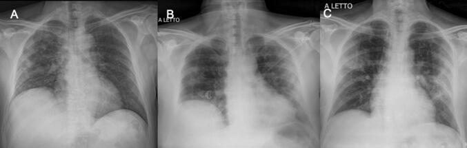
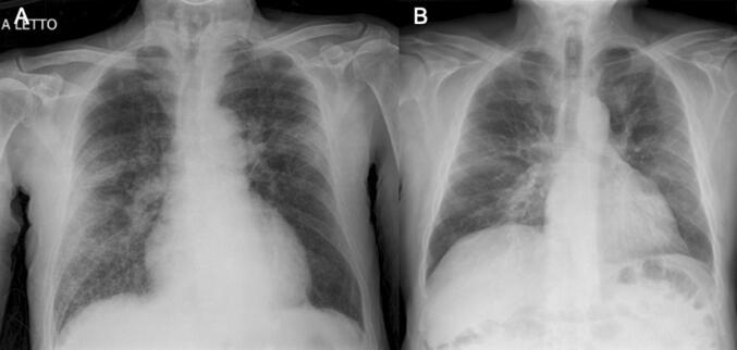
Conclusion: In COVID-19, CXR shows patchy or diffuse reticular-nodular opacities and consolidation, with basal, peripheral and bilateral predominance. In our experience, baseline CXR had a sensitivity of 68.1%. The RALE score can be used in the emergency setting as a quantitative method of the extent of SARS-CoV-2 pneumonia, correlating with an increased risk of ICU admission.
Keywords: COVID-19; Chest radiography; Coronavirus; Diagnostic imaging; Infection.
Conflict of interest statement
The authors declare that they have no conflict of interest related to the publication of this article.
Figures


References
MeSH terms
LinkOut - more resources
Full Text Sources
Miscellaneous
