Case Reports
. 2020 Jun 11;382(24):2337-2343.
doi: 10.1056/NEJMoa2000024.

Disseminated Coccidioidomycosis Treated with Interferon-γ and Dupilumab

Affiliations
Case Reports

Disseminated Coccidioidomycosis Treated with Interferon-γ and Dupilumab

Monica Tsai et al. N Engl J Med. .

Abstract

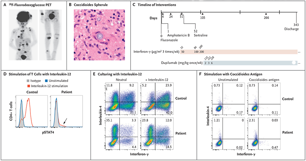
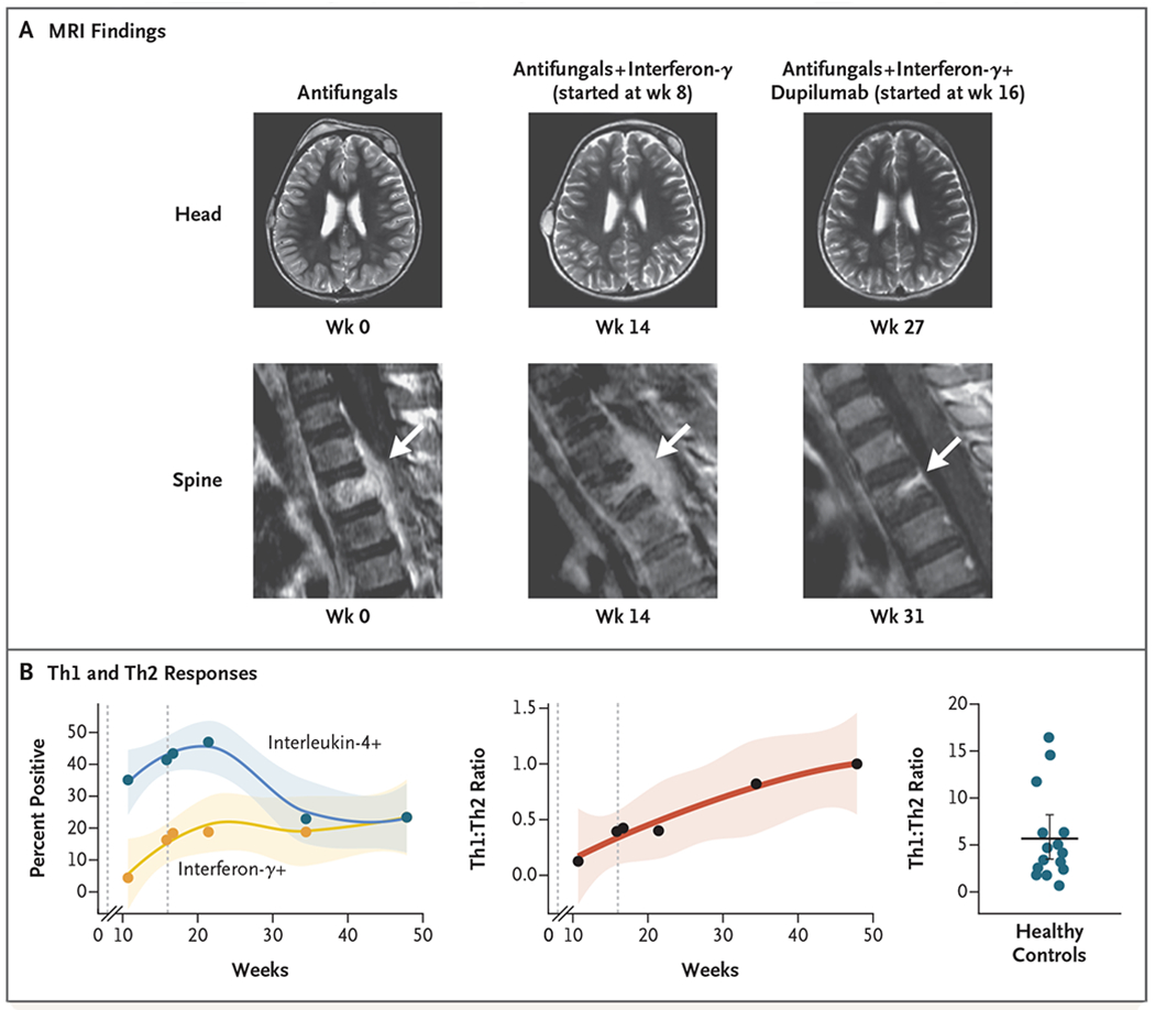
We describe a case of life-threatening disseminated coccidioidomycosis in a previously healthy child. Like most patients with disseminated coccidioidomycosis, this child had no genomic evidence of any known, rare immune disease. However, comprehensive immunologic testing showed exaggerated production of interleukin-4 and reduced production of interferon-γ. Supplementation of antifungal agents with interferon-γ treatment slowed disease progression, and the addition of interleukin-4 and interleukin-13 blockade with dupilumab resulted in rapid resolution of the patient's clinical symptoms. This report shows that blocking of type 2 immune responses can treat infection. This immunomodulatory approach could be used to enhance immune clearance of refractory fungal, mycobacterial, and viral infections. (Supported by the Jeffrey Modell Foundation and the National Institutes of Health.).


Figures

Figure 1.
Figure 1.. A Case of Disseminated Coccidioidomycosis Characterized by Defective Interleukin-12 Signaling and Th1 Response.
Panel A shows an 18F-fluorodeoxyglucose positron-emission tomographic scan showing disseminated infection with multiple lesions of the spine, clavicle, ribs, paratracheal lymph nodes, right distal radius, and right leg. Panel B shows a coccidioides spherule obtained from surgical biopsy of a scalp lesion. Panel C shows the timeline of interventions in our patient. Initial treatment included fluconazole and liposomal amphotericin B, and sertraline was added at day 52 after admission. Treatment with subcutaneous interferon-γ was also started on day 52, and treatment with dupilumab was started on day 114. Triangles represent major debridement surgical procedures. Doses of interferon-γ and dupilumab are indicated in the shaded bars; numbers above the bars are days after admission. Panel D shows stimulation of helper T cells with interleukin-12, which led to a poor phosphorylated STAT4 (pSTAT4) response; however, the loss of function was not absolute (arrow). Panel E shows intracellular cytokine staining of CD4+ T-cell effectors generated in neutral conditions and stimulated with phorbol myristate acetate (PMA) and ionomycin. Interleukin-4 production was greatly enhanced relative to interferon-γ production in the patient as compared with a control. A normal response was only partially restored by culturing in type 1 helper T (Th1) cell conditions (i.e., with interleukin-12). Panel F shows stimulation of peripheral-blood mononuclear cells with T27K coccidioidal antigen, which led to increased production of interleukin-4 over interferon-γ in helper T cells.
Figure 2.
Figure 2.. Resolution of Disseminated Coccidioidomycosis after Treatment with Interferon-γ and Dupilumab.
Panel A shows magnetic resonance imaging (MRI) of the head and spine at baseline and during treatment. A spinal lesion is indicated by the arrow. Panel B shows the percentage of CD4+ T cells producing interferon-γ (Th1 cells) or interleukin-4 (type 2 helper T [Th2] cells) (left) and their ratio (center) over time. The ratio does not include doublepositive (i.e., positive for interferon-γ and interleukin-4) cells. The first dashed line represents the initiation of interferon-γ treatment, and the second dashed line represents the initiation of dupilumab treatment. Shading indicates the 95% confidence interval. For comparison, the Th1:Th2 ratio for 15 healthy controls is shown on the right. The horizontal line indicates the bootstrapped mean, and the I bar indicates the 95% confidence interval.

References

    1. Galgiani JN, Ampel NM, Blair JE, et al. Coccidioidomycosis. Clin Infect Dis 2005;41:1217–23. - PubMed
    1. Odio CD, Marciano BE, Galgiani JN, Holland SM. Risk factors for disseminated coccidioidomycosis, United States. Emerg Infect Dis 2017;23:308–11. - PMC - PubMed
    1. Dewsnup DH, Galgiani JN, Graybill JR, et al. Is it ever safe to stop azole therapy for Coccidioides immitis meningitis? Ann Intern Med 1996;124:305–10. - PubMed
    1. Pfaller MA, Diekema DJ. Epidemiology of invasive mycoses in North America. Crit Rev MicrobioI 2010;36:1–53. - PubMed
    1. Cox RA, Baker BS, Stevens DA. Specificity of immunogIobuIin E in coccidioidomycosis and correIation with disease invoIvement. Infect Immun 1982;37:609–16. - PMC - PubMed

Publication types

MeSH terms