. 2020 Oct:255:120149.
doi: 10.1016/j.biomaterials.2020.120149. Epub 2020 May 28.

Biomimetic and estrogenic fibers promote tissue repair in mice and human skin via estrogen receptor β

Affiliations

Biomimetic and estrogenic fibers promote tissue repair in mice and human skin via estrogen receptor β

Seungkuk Ahn et al. Biomaterials. 2020 Oct.

Abstract

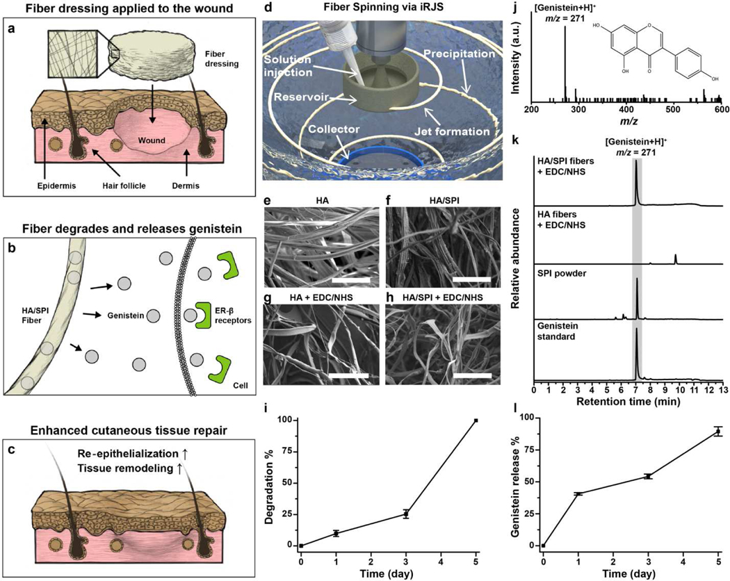
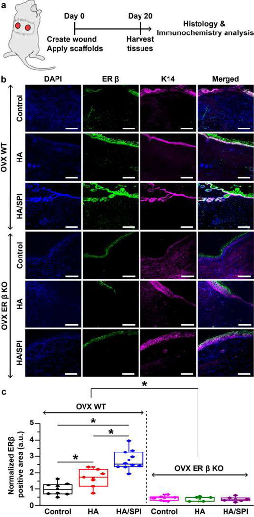
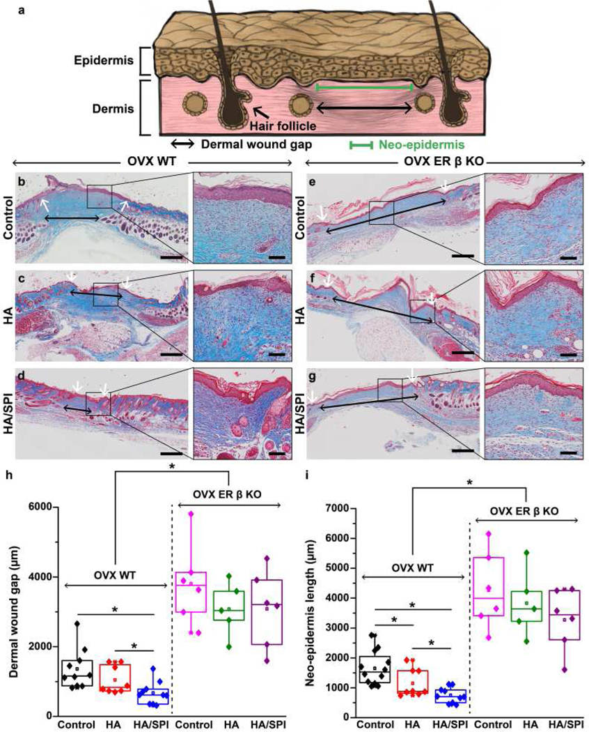
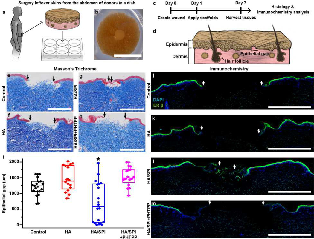
The dynamic changes in estrogen levels throughout aging and during the menstrual cycle influence wound healing. Elevated estrogen levels during the pre-ovulation phase accelerate tissue repair, whereas reduced estrogen levels in post-menopausal women lead to slow healing. Although previous reports have shown that estrogen may potentiate healing by triggering the estrogen receptor (ER)-β signaling pathway, its binding to ER-α has been associated with severe collateral effects and has therefore limited its use as a therapeutic agent. To this end, soy phytoestrogens, which preferentially bind to the ER-β, are currently being explored as a safer therapeutic alternative to estrogen. However, the development and evaluation of phytoestrogen-based materials as local ER-β modulators remains largely unexplored. Here, we engineered biomimetic and estrogenic nanofiber wound dressings built from soy protein isolate (SPI) and hyaluronic acid (HA) using immersion rotary jet spinning. These engineered scaffolds were shown to successfully recapitulate the native dermal architecture, while delivering an ER-β-triggering phytoestrogen (genistein). When tested in ovariectomized mouse and ex vivo human skin tissues, HA/SPI scaffolds outperformed controls (no treatment or HA only scaffolds) towards promoting cutaneous tissue repair. These improved healing outcomes were prevented when the ER-β pathway was genetically or chemically inhibited. Our findings suggest that estrogenic fibrous scaffolds facilitate skin repair by ER-β activation.

Keywords: Estrogen receptor β; Immersion rotary jet spinning; Nanofiber; Soy phytoestrogen; Wound healing.


Conflict of interest statement

Conflict of interests

The authors declare no competing financial interests.

Figures

Fig. 1.
Fig. 1.. Fabrication and characterization of genistein-containing fibers.
a–c, Schematic of proposed role of HA/SPI fiber scaffolds for activating the ER-β pathway in cutaneous wounds. (a) The engineered scaffolds are locally applied to the wound site. (b) During healing processes, the fibers degrade over time and continuously release genistein, an ER-β modulating molecule. The genistein released from the scaffolds binds to the ER-β, (c) promoting skin tissue repair. d, Schematic of the iRJS machine. e–h, SEM images of the engineered fibers. The scales are 50 μm. i, Scaffold degradation kinetics were characterized by measuring the weight loss of crosslinked HA/SPI scaffolds in PBS solution. j–l, LC-MS analysis of genistein content in scaffolds. (j) Full MS spectra of genistein showing the major peak at m/z 271 (inset: molecular structure of genistein). (k) LC-MS spectra of samples, recorded in SIM mode. The gray area indicates the genistein-specific peaks (retention time: 7 min). (l) Genistein release kinetics of crosslinked HA/SPI scaffolds in PBS solution. For statistical analysis, n=3 and errors bars=standard error of mean (SEM) in i and l.
Fig. 2.
Fig. 2.. ER-β expression in mouse skin at day 20 post-injury.
a, Schematic illustration of the experimental timeline. b, Immunofluorescence images of day 20 post-injury mouse skin stained with DAPI (for nuclei), anti-ER-β, and anti-K14 (for keratinocytes) antibodies. Scales are 100 μm. c, ER-β expression analysis. For statistical analysis, *p<0.05, for OVX WT mice, n=8 for the control group, n=7 for the HA group, n=10 for the HA/SPI group. For OVX ER-β KO mice, n=6 for the control and HA/SPI group, n=5 for the HA group. Box plots with all data points overlapping show data where edges, middle bars, and whiskers indicated 25th and 75th percentiles, median, 5th and 95th percentiles, respectively.
Fig. 3.
Fig. 3.. In vivo mouse wound healing and histological studies.
a, Schematic illustration of the wound healing evaluation parameters. b–g, Masson’s trichrome images of day 20 post-injury wounds treated with control (no treatment), HA scaffolds, and HA/SPI scaffolds for (b–d) OVX WT and (e–g) OVX ER-β KO mice with the zoom-in images of healed wounds on the right panels. Black and white arrows indicate dermal wound gaps and the edges of neo-epidermis, respectively. Scales of left and right panels are 500 and 100 μm, respectively. h–i, Quantitative analysis of skin tissue repair (h, dermal wound gap and i, neo-epidermis length) at day 20 post-injury. For statistical analysis, *p<0.05, For OVX WT mice, n=10 for the control and HA/SPI group, n=8 for the HA group. For OVX ER-β KO mice, n=6 for the control and HA/SPI group, n=5 for the HA group. Box plots with all data points overlapping show data where edges, middle bars, and whiskers indicated 25th and 75th percentiles, median, 5th and 95th percentiles, respectively.
Fig. 4.
Fig. 4.. Ex vivo human skin wound healing via ER-β.
a, Schematic illustration of ex vivo human skin models. b–c, (b) Photograph of a wounded human skin biopsy on day 1 prior to scaffold application with (c) experimental timeline. d, Schematic illustration of the reepithelialization analysis. e–h, Masson’s trichrome images of day 7 post-injury tissues. The black arrows indicate the edges of the new epithelial tongues. Scales are 1 mm. i, Assessment of re-epithelialization analysis at day 7 post-injury. For statistical analysis, *p<0.05, n=9 for the control group, n=8 for the HA group, n=9 for the HA/SPI group, and n=8 for the HA/SPI+PHTPP group, 2 sections per tissue from 2 different patients. Box plots with all data points overlapping show data where edges, middle bars, and whiskers indicated 25th and 75th percentiles, median, 5th and 95th percentiles, respectively. j–m, Immunofluorescence images of day 7 post-injury tissues stained with DAPI (for nuclei) and anti-ER-β antibody. The white arrows indicate the edges of the new epithelial tongues. Scales are 1 mm.

Similar articles

Cited by

References

    1. Hall G, Phillips TJ, Estrogen and skin: the effects of estrogen, menopause, and hormone replacement therapy on the skin, J. Am. Acad. Dermatol 53(4) (2005) 555–568. - PubMed
    1. Brisken C, Progesterone signalling in breast cancer: a neglected hormone coming into the limelight, Nat. Rev. Cancer 13(6) (2013) 385. - PubMed
    1. Lopez MM, Castillo AC, Kaltwasser K, Phillips LG, Moliver CL, Surgical timing and the menstrual cycle affect wound healing in young breast reduction patients, Plast. Reconstr. Surg 137(2) (2016) 406–410. - PubMed
    1. LeBlanc ES, Janowsky J, Chan BK, Nelson HD, Hormone replacement therapy and cognition: systematic review and meta-analysis, Jama 285(11) (2001) 1489–1499. - PubMed
    1. Ashcroft GS, Greenwell-Wild T, Horan MA, Wahl SM, Ferguson MW, Topical estrogen accelerates cutaneous wound healing in aged humans associated with an altered inflammatory response, Am J Pathol 155(4) (1999) 1137–1146. - PMC - PubMed

Publication types