Morphological Evaluation of Bone by CT to Determine Primary Stability-Clinical Study
- PMID: 32521622
- PMCID: PMC7321591
- DOI: 10.3390/ma13112605
Morphological Evaluation of Bone by CT to Determine Primary Stability-Clinical Study
Abstract
Background: Primary stability is an important prognostic factor for dental implant therapy. In the present study, we evaluate the relationship between implant stability evaluation findings by the use of an implant stability quotient (ISQ), an index for primary stability, and a morphological evaluation of bone by preoperative computed tomography (CT).
Subjects and methods: We analyzed 98 patients who underwent implant placement surgery in this retrospective study. For all 247 implants, the correlations of the ISQ value with cortical bone thickness, cortical bone CT value, cancellous bone CT value, insertion torque value, implant diameter, and implant length were examined.
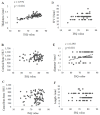
Results: 1. Factors affecting ISQ values in all cases: It was revealed that there were significant associations between the cortical bone thickness and cancellous bone CT values with ISQ by multiple regression analysis. 2. It was revealed that there was a significant correlation between cortical bone thickness and cancellous bone CT values with ISQ by multiple regression analysis in the upper jaw. 3. It was indicated that there was a significant association between cortical bone thickness and implant diameter with ISQ by multiple regression analysis in the lower jaw.
Conclusion: We concluded that analysis of the correlation of the ISQ value with cortical bone thickness and values obtained in preoperative CT imaging were useful preoperative evaluations for obtaining implant stability.
Keywords: computed tomography value; dental implants; implant stability quotient; primary stability.
Conflict of interest statement
The authors declare no conflict of interest.
Figures


Similar articles
-
The relationship between the bone characters obtained by CBCT and primary stability of the implants.Int J Implant Dent. 2015 Dec;1(1):3. doi: 10.1186/s40729-014-0003-x. Epub 2015 Feb 12. Int J Implant Dent. 2015. PMID: 27747625 Free PMC article.
-
Influence of cortical bone anchorage on the primary stability of dental implants.Oral Maxillofac Surg. 2018 Sep;22(3):297-301. doi: 10.1007/s10006-018-0705-y. Epub 2018 Jun 6. Oral Maxillofac Surg. 2018. PMID: 29876688
-
Relationship between the CT Value and Cortical Bone Thickness at Implant Recipient Sites and Primary Implant Stability with Comparison of Different Implant Types.Clin Implant Dent Relat Res. 2016 Feb;18(1):107-16. doi: 10.1111/cid.12261. Epub 2014 Sep 2. Clin Implant Dent Relat Res. 2016. PMID: 25181581
-
The Interrelation between Cortical Bone Thickness and Primary and Secondary Dental Implant Stability: a Systematic Review.J Oral Maxillofac Res. 2024 Dec 31;15(4):e2. doi: 10.5037/jomr.2024.15402. eCollection 2024 Oct-Dec. J Oral Maxillofac Res. 2024. PMID: 40017687 Free PMC article. Review.
-
The clinical significance of implant stability quotient (ISQ) measurements: A literature review.J Oral Biol Craniofac Res. 2020 Oct-Dec;10(4):629-638. doi: 10.1016/j.jobcr.2020.07.004. Epub 2020 Aug 14. J Oral Biol Craniofac Res. 2020. PMID: 32983857 Free PMC article. Review.
Cited by
-
Reducing Healing Period with DDM/rhBMP-2 Grafting for Early Loading in Dental Implant Surgery.Tissue Eng Regen Med. 2025 Feb;22(2):261-271. doi: 10.1007/s13770-024-00689-3. Epub 2025 Jan 18. Tissue Eng Regen Med. 2025. PMID: 39825990 Free PMC article.
References
-
- Japanese Society for Oral and Maxillofacial Radiology . Guidelines for the Diagnostic Imaging in Dental Implant Treatment. 2nd ed. Vol. 2. Niigata University Graduate School of Medical and Dental Studies; Niigata City, Japan: 2008. pp. 1–30.
-
- Widmann G., Bale R.J. Accuracy in computer-aided implant surgery: A review. Int. J. Oral Maxillofac. Implants. 2006;21:305–313. - PubMed
LinkOut - more resources
Full Text Sources

