Review
. 2020 Jun 8;12(6):1493.
doi: 10.3390/cancers12061493.

Diffusion-Weighted Imaging in Oncology: An Update

Affiliations
Review

Diffusion-Weighted Imaging in Oncology: An Update

Carmelo Messina et al. Cancers (Basel). .

Abstract

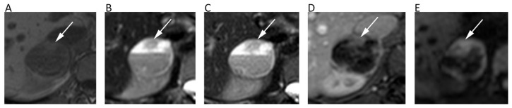
To date, diffusion weighted imaging (DWI) is included in routine magnetic resonance imaging (MRI) protocols for several cancers. The real additive role of DWI lies in the "functional" information obtained by probing the free diffusivity of water molecules into intra and inter-cellular spaces that in tumors mainly depend on cellularity. Although DWI has not gained much space in some oncologic scenarios, this non-invasive tool is routinely used in clinical practice and still remains a hot research topic: it has been tested in almost all cancers to differentiate malignant from benign lesions, to distinguish different malignant histotypes or tumor grades, to predict and/or assess treatment responses, and to identify residual or recurrent tumors in follow-up examinations. In this review, we provide an up-to-date overview on the application of DWI in oncology.

Keywords: apparent diffusion coefficient; cancer imaging; diffusion weighted imaging; magnetic resonance imaging; oncologic imaging.


Conflict of interest statement

The authors declare no conflict of interest.

Figures

Figure 1
Figure 1
Diffusion weighted imaging (DWI) b = 1000 images (A,D,G), corresponding apparent diffusion coefficient (ADC) maps (B,E,H), and contrast–enhanced T1 w images (C,F,I) of patients with histologically proven primary central nervous system lymphoma (AC), cerebral abscess (DF), and glioblastoma (GI). Mean ADC values in lymphoma (0.648 × 10⁻³ mm2/s) and in the central portion of the abscess (0.510 × 10⁻³ mm2/s) were lower than in glioblastoma. In particular, mean ADC values were higher in the necrotic core of the GBM (1.082 × 10⁻³ mm2/s) in comparison to the abscess.
Figure 2
Figure 2
MRI scans of a 65-year-old male patient with oropharyngeal squamous cell carcinoma and lymph nodal metastases. Contrast-enhanced T1w (A), high b-value DWI (B), and corresponding ADC map (C) before chemotherapy show round enlarged right cervical lymph nodes (arrows) with restricted pattern of diffusion (mean ADC: 0.891 × 10⁻³ mm2/s). The same images after treatment (DF) show the good response to chemotherapy with normal size and unrestricted pattern of diffusion of the cervical lymph nodes (curved arrows).
Figure 3
Figure 3
Chest MRI of a 62-year-old male patient with lung carcinoma. The coronal short tau inversion recovery (STIR) (A), coronal reconstruction of b 1000 DWI image (B), axial post-contrast T1w image (C), axial b = 1000 DWI image (D), and corresponding ADC map (E) show a large tumor in the left lung (arrows) with an restricted pattern of diffusion. Note the central necrotic areas (asterisks) presenting fluid signal intensity on STIR (A), absent contrast enhancement (C) and unrestricted pattern of diffusion (D,E) with mean ADC of 2.168 × 10⁻³ mm2/s. Conversely, the solid component of the tumor shows strong contrast enhancement and very low ADC values (0.818 × 10⁻³ mm2/s).
Figure 4
Figure 4
Breast MRI of a 42-year-old female patient with right breast carcinoma. Axial IDEAL fat only (A) and IDEAL water only (B) images show a right breast carcinoma (arrows) with strong enhancement on post-contrast T1w image (C) and high signal intensity on b = 900 DWI image (D).
Figure 5
Figure 5
Liver MRI of a 54-year-old female patient with histologically proven hepatocellular carcinoma (HCC) on her healthy liver. Axial T2w (A) and pre-contrast fat-suppressed 3D-gradient-echo T1w (B) images show a large mass with strong contrast enhancement on the arterial phase (C), wash-out and rim enhancement on the portal phase (D), high signal intensity on b = 800 DWI image (E), and low ADC values (mean ADC: 1.018 × 10⁻³ mm2/s) on the corresponding ADC map (F).
Figure 6
Figure 6
Upper abdomen MRI of a patient with G2 pancreatic neuroendocrine neoplasm. A heterogeneous hypervascular lesion (arrows) with well-defined margins can be seen on the axial arterial phase image (A). The lesion is clearly hyperintense on b = 800 DWI (B), with low ADC values on the corresponding ADC map (C). IVIM maps show that the tumor has low values of fast diffusion (D) Dp, but relatively preserved perfusion fraction (E) Fp, consistent with a neuroendocrine neoplasm.
Figure 7
Figure 7
MRI scans of a 66-year-old female patient with rectal cancer at the initial diagnosis and two months later after neoadjuvant chemotherapy. Axial T2w image (A) and axial post-contrast fat-suppressed 3D GRE T1w image (B) show the tumor along the posterior rectal wall (arrows) with high signal intensity on the axial b = 600 DWI image (C) and low ADC (mean ADC: 0.651 × 10⁻³ mm2/s) on the corresponding ADC map (D). Post-treatment axial T2w image (E) shows the reduction in size of the lesion (arrowheads) with decrease of contrast enhancement (F) and slight increase of ADC values (G,H).
Figure 8
Figure 8
A cervical cancer without parametrial invasion in a 70-year-old woman. The lesion (arrows) is slightly hyperintense compared to normal cervical stroma on sagittal (A) and axial (B) T2w, whereas it is highly hyperintense on b = 800 DWI (C) and hypointense on the corresponding ADC map (D), with moderate contrast enhancement on fat-suppressed 3D-GRE T1w (E).
Figure 9
Figure 9
A 28-year-old male patient with pheochromocytoma of the right adrenal gland. Axial in-phase T1w (A), axial T2w (B), and axial fat-suppressed T2w (C) images show a right adrenal cystic lesion with fluid-fluid level. Axial post-contrast fat-suppressed 3D GRE T1w (D) and b = 600 DWI (E) allow one to better depict the solid component of the lesion consisting of the peripheral wall and the anterior nodular portion (arrows), with no restricted pattern of diffusion in central cystic part.
Figure 10
Figure 10
A 68-year-old male patient with prostate cancer. Axial T2w (A) and post-contrast fat-suppressed T1w (B) images show a hypointense nodule in the left peripheral zone (arrows) with high signal intensity on b = 1000 DWI image (C) and restricted pattern of diffusion on the corresponding ADC map (D).
Figure 11
Figure 11
Whole-body MRI of a 58-year-old female patient with non-Hodgkin lymphoma. Whole-body T1w (A) and coronal MIP grey-scale inverted DWI (B) show multiple nodal and bone locations of disease. Note the periaortic lymph nodal locations (arrows) with moderate contrast enhancement on axial fat-suppressed 3D GRE T1w (C) and restricted pattern of diffusion on axial b = 800 DWI (D) and corresponding ADC map (E, mean ADC: 0.761 × 10⁻³ mm2/s).
Figure 12
Figure 12
The left femur G3 chondrosarcoma (AE) of a 76-year-old male patient. Axial T2w image (A) shows a hyperintense bone lesion with a large posterior soft tissue mass presenting poor contrast enhancement on fat-suppressed T1w image (B). The lesion shows high signal on b = 0 DWI image (C), decreased signal on b = 1000 DWI image (D), and unrestricted pattern of diffusion on the corresponding ADC map (E, mean ADC: 2.303 × 10⁻³ mm2/s). The Ewing sarcoma (FJ) of the left iliac bone of a 31-year-old male patient. The tumor presents as an esofitic bone lesion with intermediate signal on axial T2w image (F) and high signal on axial STIR image (G). The lesion shows intermediate-high signal on b = 0 DWI image (H), higher signal on b = 1000 DWI image (I), and restricted pattern of diffusion on the corresponding ADC map (J, mean ADC: 0.899 × 10⁻³ mm2/s).
Figure 13
Figure 13
MR images of an intramuscular lipoma of the forearm (AD) and a perineural ganglion cyst of the leg (EG) of two different patients. The lipoma (asterisk) shows a homogeneous high signal on coronal T2w (A) and a low signal on axial fat-suppressed T1w (B), b = 800 DWI (C); the corresponding ADC map (D). Note the absence of diffusion restriction and the intrinsically low ADC values of this benign lipomatous lesion. The ganglion cyst (arrows) displays high signal intensity on coronal fat-suppressed T2w (E), axial b = 800 DWI (F), and axial ADC map (G). High b-value DWI shows high signal due to T2 shine-through effect; the ADC map confirms the absence of diffusion restriction and the cystic nature of the lesion (ADC: 2.603 × 10⁻³ mm2/s).

References

    1. Svolos P., Kousi E., Kapsalaki E., Theodorou K., Fezoulidis I., Kappas C., Tsougos I. The role of diffusion and perfusion weighted imaging in the differential diagnosis of cerebral tumors: A review and future perspectives Tumour charcterisation. Cancer Imaging. 2014;14:20. doi: 10.1186/1470-7330-14-20. - DOI - PMC - PubMed
    1. Muccio C.F., Caranci F., D’Arco F., Cerase A., De Lipsis L., Esposito G., Tedeschi E., Andreula C. Magnetic resonance features of pyogenic brain abscesses and differential diagnosis using morphological and functional imaging studies: A pictorial essay. J. Neuroradiol. 2014;41:153–167. doi: 10.1016/j.neurad.2014.05.004. - DOI - PubMed
    1. Yamasaki F., Kurisu K., Satoh K., Arita K., Sugiyama K., Ohtaki M., Takaba J., Tominaga A., Hanaya R., Yoshioka H., et al. Apparent diffusion coefficient of human brain tumors at MR imaging. Radiology. 2005;235:985–991. doi: 10.1148/radiol.2353031338. - DOI - PubMed
    1. Rosenkrantz A.B., Padhani A.R., Chenevert T.L., Koh D.-M., De Keyzer F., Taouli B., Le Bihan D. Body diffusion kurtosis imaging: Basic principles, applications, and considerations for clinical practice. J. Magn. Reson. Imaging. 2015;42:1190–1202. doi: 10.1002/jmri.24985. - DOI - PubMed
    1. Lee E.J., terBrugge K., Mikulis D., Choi D.S., Bae J.M., Lee S.K., Moon S.Y. Diagnostic value of peritumoral minimum apparent diffusion coefficient for differentiation of glioblastoma multiforme from solitary metastatic lesions. AJR Am. J. Roentgenol. 2011;196:71–76. doi: 10.2214/AJR.10.4752. - DOI - PubMed