Improved Outcome Prediction for Appendiceal Pseudomyxoma Peritonei by Integration of Cancer Cell and Stromal Transcriptional Profiles
- PMID: 32521738
- PMCID: PMC7352410
- DOI: 10.3390/cancers12061495
Improved Outcome Prediction for Appendiceal Pseudomyxoma Peritonei by Integration of Cancer Cell and Stromal Transcriptional Profiles
Abstract
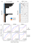
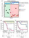
In recent years, cytoreductive surgery (CRS) and hyperthermic intraperitoneal chemotherapy (HIPEC) have substantially improved the clinical outcome of pseudomyxoma peritonei (PMP) originating from mucinous appendiceal cancer. However, current histopathological grading of appendiceal PMP frequently fails in predicting disease outcome. We recently observed that the integration of cancer cell transcriptional traits with those of cancer-associated fibroblasts (CAFs) improves prognostic prediction for tumors of the large intestine. We therefore generated global expression profiles on a consecutive series of 24 PMP patients treated with CRS plus HIPEC. Multiple lesions were profiled for nine patients. We then used expression data to stratify the samples by a previously published "high-risk appendiceal cancer" (HRAC) signature and by a CAF signature that we previously developed for colorectal cancer, or by a combination of both. The prognostic value of the HRAC signature was confirmed in our cohort and further improved by integration of the CAF signature. Classification of cases profiled for multiple lesions revealed the existence of outlier samples and highlighted the need of profiling multiple PMP lesions to select representative samples for optimal performances. The integrated predictor was subsequently validated in an independent PMP cohort. These results provide new insights into PMP biology, revealing a previously unrecognized prognostic role of the stromal component and supporting integration of standard pathological grade with the HRAC and CAF transcriptional signatures to better predict disease outcome.
Keywords: appendiceal cancer; cancer-associated fibroblasts.; gene expression profiling; peritoneal carcinosis; prognostic signatures; pseudomyxoma peritonei; tumor stroma.
Conflict of interest statement
The authors declare no conflicts of interest.
Figures


Similar articles
-
Cytoreductive surgery (CRS) and hyperthermic intraperitoneal chemotherapy (HIPEC) in pseudomyxoma peritonei of appendiceal origin: result of a single centre study.Updates Surg. 2020 Dec;72(4):1207-1212. doi: 10.1007/s13304-020-00788-5. Epub 2020 May 14. Updates Surg. 2020. PMID: 32410159
-
The Role of Hyperthermic Intraperitoneal Chemotherapy in Pseudomyxoma Peritonei After Cytoreductive Surgery.JAMA Surg. 2021 Mar 1;156(3):e206363. doi: 10.1001/jamasurg.2020.6363. Epub 2021 Mar 10. JAMA Surg. 2021. PMID: 33502455 Free PMC article.
-
Reclassification of Appendiceal Mucinous Neoplasms and Associated Pseudomyxoma Peritonei According to the Peritoneal Surface Oncology Group International Consensus: Clinicopathological Reflections of a Two-Center Cohort Study.Ann Surg Oncol. 2024 Dec;31(13):8572-8584. doi: 10.1245/s10434-024-16254-0. Epub 2024 Sep 26. Ann Surg Oncol. 2024. PMID: 39327362
-
Cytoreductive surgery combined with hyperthermic intraperitoneal chemotherapy for Pseudomyxoma peritonei originating from ovarian teratomas: A single-center case series and literature review.Eur J Obstet Gynecol Reprod Biol. 2025 May;309:107-112. doi: 10.1016/j.ejogrb.2025.03.043. Epub 2025 Mar 18. Eur J Obstet Gynecol Reprod Biol. 2025. PMID: 40117798 Review.
-
Pathology of Mucinous Appendiceal Tumors and Pseudomyxoma Peritonei.Indian J Surg Oncol. 2016 Jun;7(2):258-67. doi: 10.1007/s13193-016-0516-2. Epub 2016 Mar 19. Indian J Surg Oncol. 2016. PMID: 27065718 Free PMC article. Review.
Cited by
-
Tumor-stroma ratio as a new prognosticator for pseudomyxoma peritonei: a comprehensive clinicopathological and immunohistochemical study.Diagn Pathol. 2021 Dec 13;16(1):116. doi: 10.1186/s13000-021-01177-1. Diagn Pathol. 2021. PMID: 34895284 Free PMC article.
-
Clinicopathological Characteristics and Prognostic Prediction in Pseudomyxoma Peritonei Originating From Mucinous Ovarian Cancer.Front Oncol. 2021 Apr 21;11:641053. doi: 10.3389/fonc.2021.641053. eCollection 2021. Front Oncol. 2021. PMID: 33968739 Free PMC article.
-
A Review of Pseudomyxoma Peritonei: Insights Into Diagnosis, Management, and Prognosis.Cureus. 2024 May 28;16(5):e61244. doi: 10.7759/cureus.61244. eCollection 2024 May. Cureus. 2024. PMID: 38939264 Free PMC article. Review.
-
A nomogram prediction model of pseudomyxoma peritonei established based on new prognostic factors of HE stained pathological images analysis.Cancer Med. 2024 Mar;13(6):e7101. doi: 10.1002/cam4.7101. Cancer Med. 2024. PMID: 38506243 Free PMC article.
-
Distinct gene signatures define the epithelial cell features of mucinous appendiceal neoplasms and pseudomyxoma metastases.Front Genet. 2025 Feb 13;16:1536982. doi: 10.3389/fgene.2025.1536982. eCollection 2025. Front Genet. 2025. PMID: 40018643 Free PMC article.
References
-
- Sugarbaker P.H., Ronnett B.M., Archer A., Averbach A.M., Bland R., Chang D., Dalton R.R., Ettinghausen S.E., Jacquet P., Jelinek J., et al. Pseudomyxoma peritonei syndrome. Adv. Surg. 1996;30:233. - PubMed
Grants and funding
LinkOut - more resources
Full Text Sources
Molecular Biology Databases

