

Mycobacterium tuberculosis Rv0341 Promotes Mycobacterium Survival in In Vitro Hostile Environments and within Macrophages and Induces Cytokines Expression
- PMID: 32521796
- PMCID: PMC7350357
- DOI: 10.3390/pathogens9060454
Mycobacterium tuberculosis Rv0341 Promotes Mycobacterium Survival in In Vitro Hostile Environments and within Macrophages and Induces Cytokines Expression
Abstract
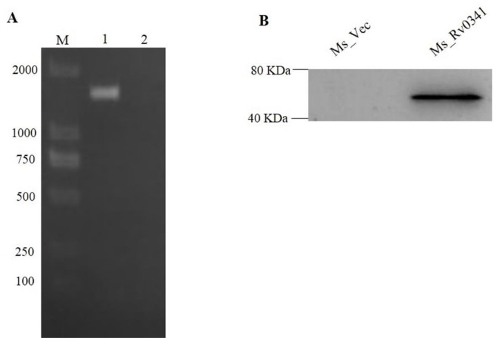
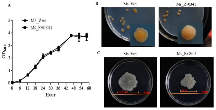
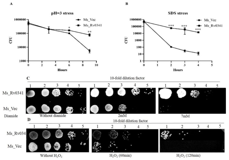
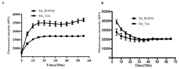
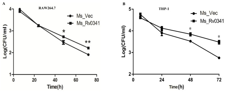
Mycobacterium tuberculosis represents an ancient deadly human pathogen that can survive and multiply within macrophages. The effectors are key players for the successful pathogenesis of this bacterium. M. tuberculosis open reading frame (ORF) Rv0341, a pathogenic mycobacteria-specific gene, was found to be upregulated in macrophages isolated from human tuberculosis granuloma and inside the macrophages during in vitro infection by M. tuberculosis. To understand the exact role of this gene, we expressed the Rv0341 gene in M. smegmatis, which is a non-pathogenic Mycobacterium. We found that Rv0341 expression can alter colony morphology, reduce the sliding capability, and decrease the cell wall permeability of M. smegmatis. Furthermore, Rv0341 remarkably enhanced M. smegmatis survival within macrophages and under multiple in vitro stress conditions when compared with the control strain. Ms_Rv0341 significantly induced expression of TNF-α, IL-1β, and IL-10 compared with M. smegmatis harboring an empty vector. In summary, these data suggest that Rv0341 is one of the M. tuberculosis virulence determinants that can promote bacilli survival in harsh conditions and inside macrophages.
Keywords: Mycobacterium tuberculosis; Rv0341; cytokines; infection; macrophages; stress.
Conflict of interest statement
The authors declare no conflicts of interest.
Figures

Similar articles
-
Mycobacterium tuberculosis PPE25 and PPE26 proteins expressed in Mycobacterium smegmatis modulate cytokine secretion in mouse macrophages and enhance mycobacterial survival.Res Microbiol. 2017 Apr;168(3):234-243. doi: 10.1016/j.resmic.2016.06.004. Epub 2016 Jun 25. Res Microbiol. 2017. PMID: 27351106
-
PE17 protein from Mycobacterium tuberculosis enhances Mycobacterium smegmatis survival in macrophages and pathogenicity in mice.Microb Pathog. 2019 Jan;126:63-73. doi: 10.1016/j.micpath.2018.10.030. Epub 2018 Oct 23. Microb Pathog. 2019. PMID: 30366126
-
Rv3539 (PPE63) of Mycobacterium Tuberculosis Promotes Survival of Mycobacterium Smegmatis in Human Macrophages Cell Line via Cell Wall Modulation of Bacteria and Altering Host's Immune Response.Curr Microbiol. 2023 Jul 4;80(8):267. doi: 10.1007/s00284-023-03360-7. Curr Microbiol. 2023. PMID: 37401981
-
PPE11 of Mycobacterium tuberculosis can alter host inflammatory response and trigger cell death.Microb Pathog. 2019 Jan;126:45-55. doi: 10.1016/j.micpath.2018.10.031. Epub 2018 Oct 23. Microb Pathog. 2019. PMID: 30366125
-
Comparing the Metabolic Capabilities of Bacteria in the Mycobacterium tuberculosis Complex.Microorganisms. 2019 Jun 18;7(6):177. doi: 10.3390/microorganisms7060177. Microorganisms. 2019. PMID: 31216777 Free PMC article. Review.
Cited by
-
Cigarette Smoking as a Risk Factor for Tuberculosis in Adults: Epidemiology and Aspects of Disease Pathogenesis.Pathogens. 2024 Feb 7;13(2):151. doi: 10.3390/pathogens13020151. Pathogens. 2024. PMID: 38392889 Free PMC article. Review.
-
Mycobacterium tuberculosis Methyltransferase Rv1515c Can Suppress Host Defense Mechanisms by Modulating Immune Functions Utilizing a Multipronged Mechanism.Front Mol Biosci. 2022 Jun 24;9:906387. doi: 10.3389/fmolb.2022.906387. eCollection 2022. Front Mol Biosci. 2022. PMID: 35813825 Free PMC article.
-
Pathogenic mechanisms and etiologic aspects of Mycobacterium avium subspecies paratuberculosis as an infectious cause of cutaneous melanoma.MedComm Oncol. 2024 Jun;3(2):e72. doi: 10.1002/mog2.72. Epub 2024 May 12. MedComm Oncol. 2024. PMID: 38831791 Free PMC article.
-
Transcriptional Response of Mycobacterium tuberculosis to Cigarette Smoke Condensate.Front Microbiol. 2021 Oct 15;12:744800. doi: 10.3389/fmicb.2021.744800. eCollection 2021. Front Microbiol. 2021. PMID: 34721344 Free PMC article.
-
Involvement of Mycobacterium smegmatis small noncoding RNA B11 in triacylglycerol accumulation and altered cell wall permeability.BMC Microbiol. 2025 Mar 8;25(1):124. doi: 10.1186/s12866-025-03826-7. BMC Microbiol. 2025. PMID: 40057673 Free PMC article.
References
-
- World Health Organization . Global Tuberculosis Report 2019. World Health Organization; Geneva, Switzerland: 2019.
Grants and funding
LinkOut - more resources
Full Text Sources
