. 2020 Jun 10;18(1):87.
doi: 10.1186/s12951-020-00644-z.

X-ray induced photodynamic therapy (PDT) with a mitochondria-targeted liposome delivery system

Affiliations

X-ray induced photodynamic therapy (PDT) with a mitochondria-targeted liposome delivery system

Xuefan Gu et al. J Nanobiotechnology. .

Abstract

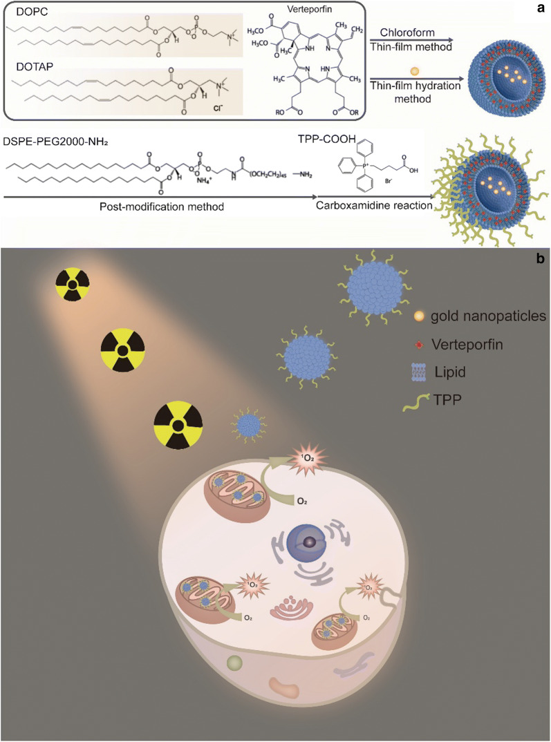
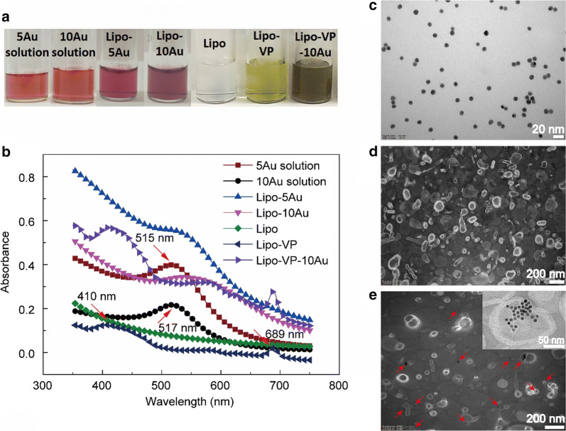
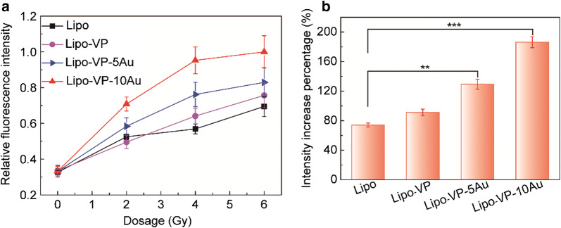
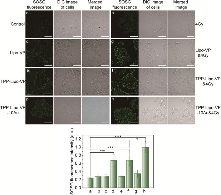
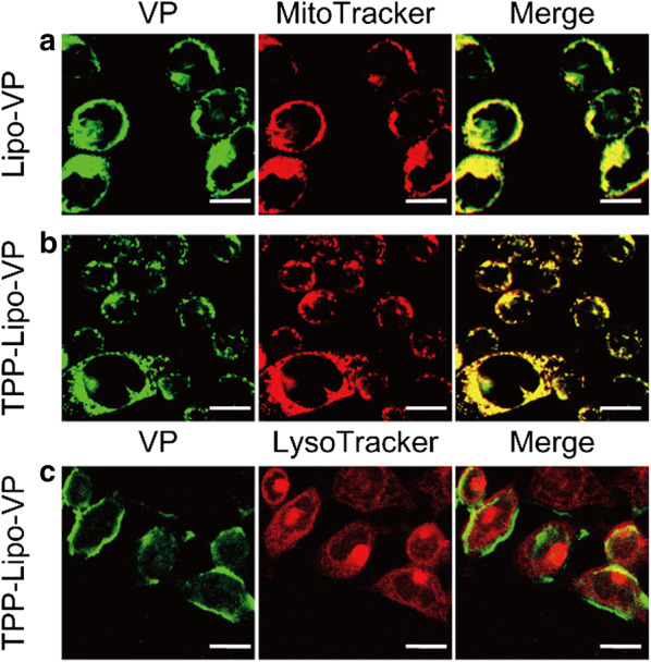
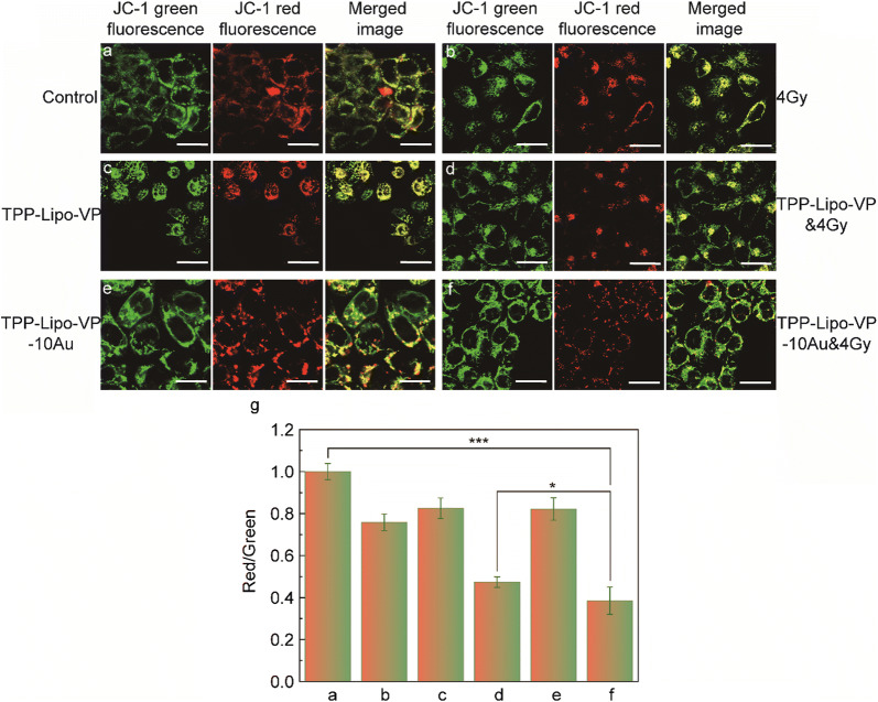
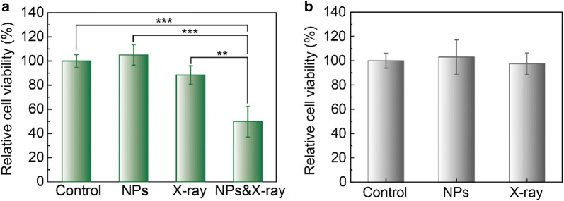
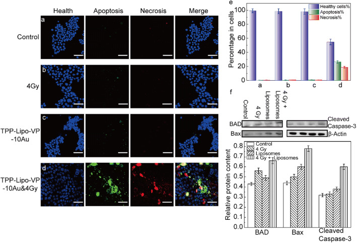
In this study, we constructed multifunctional liposomes with preferentially mitochondria-targeted feature and gold nanoparticles-assisted synergistic photodynamic therapy. We systemically investigated the in vitro X-ray triggered PDT effect of these liposomes on HCT 116 cells including the levels of singlet oxygen, mitochondrial membrane potential, cell apoptosis/necrosis and the expression of apoptosis-related proteins. The results corroborated that synchronous action of PDT and X-ray radiation enhance the generation of cytotoxic reactive oxygen species produced from the engineered liposomes, causing mitochondrial dysfunction and increasing the levels of apoptosis.

Keywords: Gold nanoparticles; Liposome; Mitochondria-targeted; Photodynamic therapy; X-ray.


Conflict of interest statement

There are no conflicts to declare.

Figures

Scheme 1
Scheme 1
The schematic illustration of a the liposome formulation and b mitochondria-targeted X-ray induced PDT via the liposome delivery system
Fig. 1
Fig. 1
Characterisation of liposomes incorporating VP and gold nanoparticles. a Photograph of liposome samples and pure gold colloidal solution. b Absorption spectra of different liposome samples and pure gold colloidal solution. Red arrows indicated typical absorption peaks of VP (~ 410 nm and ~ 689 nm), 5 nm gold nanoparticles (~ 515 nm and 10 nm gold nanoparticles ~ 517 nm). ce TEM images of 10 nm gold nanoparticles (c) pure liposomes (d) and liposomes loaded with 10 nm gold nanoparticles (e) Red arrows indicate gold nanoparticles encapsulated inside the liposomes
Fig. 2
Fig. 2
1O2 generation from different liposome samples under X-ray radiation. a Relative SOSG fluorescence intensities from liposome samples at different X-ray doses. b Percentage increase in SOSG fluorescence intensities from different liposome samples under 4 Gy X-ray radiation. Error bars show standard deviation from four measurements
Fig. 3
Fig. 3
Intracellular 1O2 production from liposomes with and without X-ray radiation. (ah) Representative confocal laser scanning microscopy images of SOSG in a HCT116 cells without any treatment, b HCT116 cells after 4 Gy X-ray irradiation, c HCT116 cells treated with Lipo-VP (250 µM) for 4 h, d HCT116 cells treated with Lipo-VP (250 µM) for 4 h and 4 Gy X-ray irradiation, e HCT116 cells treated with TPP-Lipo-VP (250 µM) for 4 h, f HCT116 cells treated with TPP-Lipo-VP (250 µM) for 4 h and 4 Gy X-ray irradiation, g HCT116 cells treated with Lipo-VP-10Au (250 µM) for 4 h and h HCT116 cells treated with Lipo-VP-10Au (250 µM) for 4 h and 4 Gy X-ray irradiation. Scale bar is 70 µm. g Quantitative analysis of 1O2 generation in HCT116 cells at different treatment conditions
Fig. 4
Fig. 4
Colocalization of Lipo-VP and TPP-Lipo-VP with the mitochondria and lysosomes. ac Representative confocal laser scanning microscopy images of VP (pseudo green colour to distinguish between VP and trackers), Mitotracker Red (red channel) and LysoTracker (red channel) in HCT116 cells after incubation with Lipo-VP (250 µM) and TPP-Lipo-VP (250 µM). Merged images of green and red channels indicated colocalization level of the liposomes and mitochondria (or lysosomes). Scale bar is 20 µm
Fig. 5
Fig. 5
Changes in JC-1 fluorescence signal (Δψm) in HCT116 cells under different treatment conditions: a the cells without any treatment, b the cells treated with 4 Gy X-ray radiation, c the cells treated with TPP-Lipo-VP (250 µM), d the cells treated with TPP-Lipo-VP (250 µM) and 4 Gy X-ray radiation, e the cells treated with TPP-Lipo-VP-10Au (250 µM) and f the cells treated with TPP-Lipo-VP-10Au (250 µM) and 4 Gy X-ray radiation. Scale bar is 20 µm. The high-membrane potential resulted in the JC-1 aggregation in the membrane, as shown by the strong red fluorescent signal, and low membrane potential caused the monodispersed release of JC-1 to the cytosol, as shown by the strong green fluorescent signal. g Red-to-green channel ratio determines the rate of membrane potential decay and cells dead
Fig. 6
Fig. 6
a In vitro cytotoxicity of TPP-Lipo-VP-10Au in HCT116 cells after X-ray irradiation. The cells were treated with X-ray alone, TPP-Lipo-VP-10Au (250 µM) alone and combined condition. b The dark cytotoxicity of TPP-Lipo-VP-10Au (250 µM) and X-ray irradiation (4 Gy) in CCD841 CoN cells
Fig. 7
Fig. 7
Apoptosis/necrosis assays performed in HCT116 cells at 24 h after the treatments. a–d Representative confocal laser scanning microscopy images of the cells after different treatments as indicated. The concentration of TPP-Lipo-VP-10Au used to treat the cells was 250 µM. Scale bar is 70 µm. e Relative changes in levels of apoptosis and necrosis upon different treatments as indicated in ad. f Relative protein expression was analysed by using ImageJ software. Protein expression was normalized by use of the loading control protein (β-actin)

References

    1. Lucky SS, Soo KC, Zhang Y. Nanoparticles in photodynamic therapy. Chem Rev. 2015;115(4):1990–2042. - PubMed
    1. Que Y, Liu Y, Tan W, et al. Enhancing photodynamic therapy efficacy by using fluorinated nanoplatform. ACS Macro Lett. 2016;5(2):168–173. - PubMed
    1. Deng K, Li C, Huang S, et al. Recent progress in near infrared light triggered photodynamic therapy. Small. 2017;13(44):1702299. - PubMed
    1. Kim S, Tachikawa T, Fujitsuka M, et al. Far-red fluorescence probe for monitoring singlet oxygen during photodynamic therapy. J Am Chem Soc. 2014;136(33):11707–11715. - PubMed
    1. Noh I, Kim HO, Choi J, et al. Co-delivery of paclitaxel and gemcitabine via CD44-targeting nanocarriers as a prodrug with synergistic antitumor activity against human biliary cancer. Biomaterials. 2015;53:763–774. - PubMed

LinkOut - more resources