Review
. 2021 Jan;141(1):84-94.e6.
doi: 10.1016/j.jid.2020.05.098. Epub 2020 Jun 6.

Notch-Mediated Generation of Monocyte-Derived Langerhans Cells: Phenotype and Function

Affiliations
Review

Notch-Mediated Generation of Monocyte-Derived Langerhans Cells: Phenotype and Function

Lydia Bellmann et al. J Invest Dermatol. 2021 Jan.

Abstract

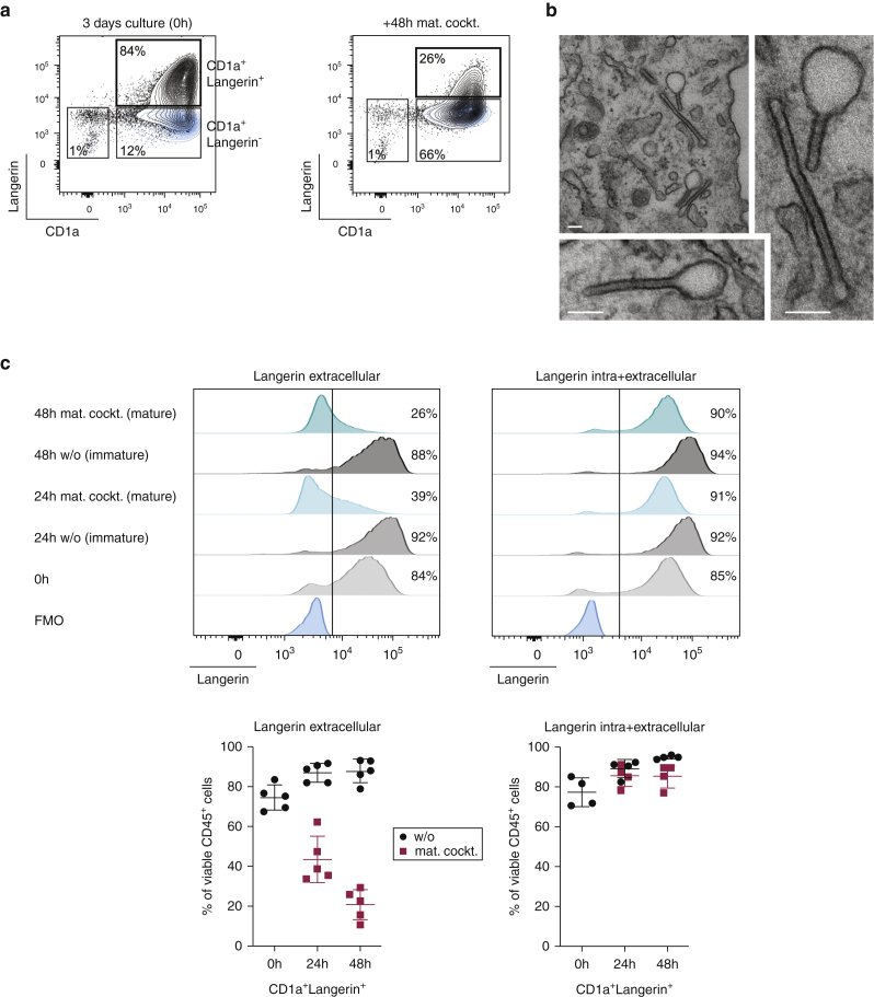
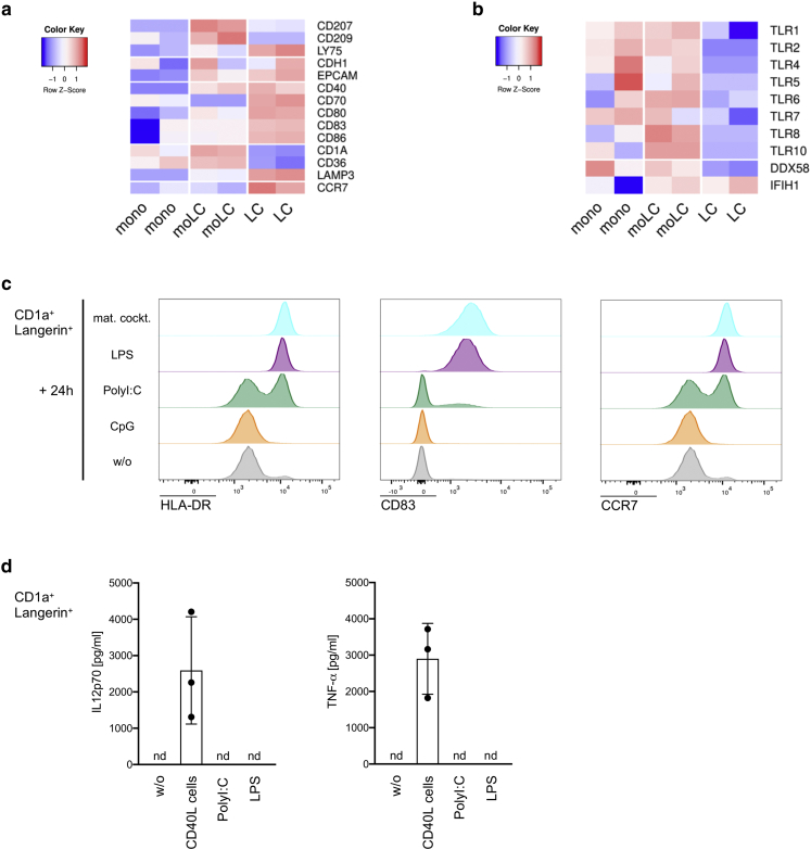
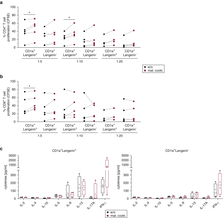
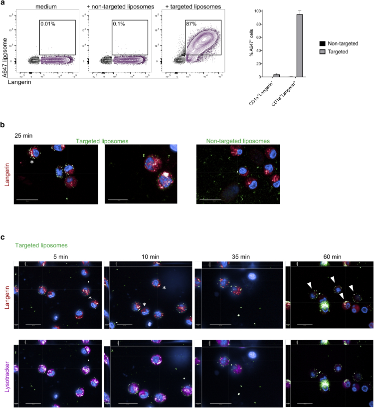
Langerhans cells (LCs) in the skin are a first line of defense against pathogens but also play an essential role in skin homeostasis. Their exclusive expression of the C-type lectin receptor Langerin makes them prominent candidates for immunotherapy. For vaccine testing, an easily accessible cell platform would be desirable as an alternative to the time-consuming purification of LCs from human skin. Here, we present such a model and demonstrate that monocytes in the presence of GM-CSF, TGF-β1, and the Notch ligand DLL4 differentiate within 3 days into CD1a+Langerin+cells containing Birbeck granules. RNA sequencing of these monocyte-derived LCs (moLCs) confirmed gene expression of LC-related molecules, pattern recognition receptors, and enhanced expression of genes involved in the antigen-presenting machinery. On the protein level, moLCs showed low expression of costimulatory molecules but prominent expression of C-type lectin receptors. MoLCs can be matured, secrete IL-12p70 and TNF-α, and stimulate proliferation and cytokine production in allogeneic CD4+ and CD8+ T cells. In regard to vaccine testing, a recently characterized glycomimetic Langerin ligand conjugated to liposomes demonstrated specific and fast internalization into moLCs. Hence, these short-term in vitro‒generated moLCs represent an interesting tool to screen LC-based vaccines in the future.


Figures

Figure 1
Figure 1
Notch ligation allows rapid differentiation of moLCs with high expression of Langerin and Birbeck granules. (a) Representative density plots showing surface Langerin and CD1a after 3-day culture of monocytes on OP9-DLL4 with TGF-β1 and GM-CSF. Mat. cockt. was added for an additional 48 hours, and cells were analyzed. (b) Representative electron microscopy of Birbeck granules from two moLCs. The right panel was enlarged from the top panel. Bars = 100 nm. (c) Representative histograms for Langerin surface (left, extracellular) and Langerin total (right, intra and extracellular) are shown on moLCs at different times before (0 h) and after maturation (mat. cockt.) or medium control (w/o). Summary graphs for five experiments show percentages of CD1a+Langerin+ cells within viable CD45+ cells. Mean ± SD. FMO, fluorescence minus one; h, hour; LC, Langerhans cell; mat. cockt, maturation cocktail; moLC, monocyte-derived LC; w/o, without.
Figure 2
Figure 2
In vitro‒generated moLCs express LC-related molecules and respond to TLR or RLR ligands. (a, b) Sorted monocytes, moLCs, and migratory skin LCs from two different donors were analyzed by RNA-seq. Heatmap depicts the normalized and relative expression (z score) of (a) LC-related genes and (b) TLR and RLR genes. (c) MoLCs were analyzed by flow cytometry after 24 hours with a cytokine mat. cockt, LPS, PolyI:C, or CpG for the expression of the maturation markers HLA-DR and CD83 and the chemokine receptor CCR7. Representative histograms of one donor (n = 2–3) are shown. (d) A total of 100,000 moLCs were cultured with 50,000 CD40L cells or TLR ligands for 24 hours, and IL-12p70 as well as TNF-α were measured in supernatants by ELISA. Mean ± SD, n = 2–3. h, hour; LC, Langerhans cell; LPS, lipopolysaccharide; mat.cockt, maturation cocktail; moLC, monocyte-derived LC; nd, not detectable; PolyI:C, polyinosinic:polycytidylic acid; RLR, RIG-I‒like receptor; TLR, toll-like receptor; RNA-seq, RNA sequencing; w/o, without.
Figure 3
Figure 3
In vitro‒generated moLCs express maturation markers and C-type lectin receptors DEC-205 and DC-SIGN. (a, b) In vitro‒generated moLCs (CD1a+Langerin+) were analyzed by flow cytometry after 3 days of culturing (0 h) or after 24 h or 48 h in the presence (mat.cockt.) or absence (w/o) of a cytokine mat. cockt. for the expression of (a) the maturation markers HLA-DR, CD83, CD80, and CD86 and (b) C-type lectin receptors DEC-205 and DC-SIGN. Summary graphs for four experiments show percentages of receptor-positive cells within CD1a+Langerin+ cells. Mean ± SD. DC, dendritic cell; FMO, fluorescence minus one; h, hour; LC, Langerhans cell; mat. cockt, maturation cocktail; moLC, monocyte-derived LC; w/o, without.
Figure 4
Figure 4
In vitro‒generated mature moLCs stimulate allogeneic T cells and induce higher Th1/Th17 cytokines. (a–c) In vitro‒generated CD1a+Langerin+ moLCs and CD1a+Langerin cells were sorted, cultured for 24 hours in the presence (mat. cockt.) or absence (w/o) of a cytokine mat. cockt. and then cocultured in different ratios with CFSE-labeled allogeneic PBLs. After 5 days, CFSE dilution was analyzed by flow cytometry for (a) CD4+ T cells and (b) CD8+ T cells for the indicated ratios. (c) Supernatants were collected on day 5 of coculture, and secretion of T cell cytokines for the 1:10 ratio was analyzed by ProcartaPlex Immunoassay. Summary graphs for four experiments are displayed as box plots with individual blood donors. CFSE, carboxyfluorescein succinimidyl ester; LC, Langerhans cell; mat. cockt, maturation cocktail; moLC, monocyte-derived LC; PBL, peripheral blood lymphocyte; Th, T helper; w/o, without.
Figure 5
Figure 5
MoLCs can be targeted with a glycomimetic Langerin ligand. (a–c) In vitro‒generated moLCs and CD1a+Langerin cells were incubated with A647-labeled liposomes coated with a Langerin ligand (targeted liposomes) or nontargeted liposomes. (a) Internalization after 1 hour at 37 °C was determined by positive A647 signal by flow cytometry. Summary graph for four experiments is shown; Mean ± SD. (b, c) Binding and internalization were followed by Operetta High-Content Imaging System for (b) 25 minutes or (c) the indicated time periods. Liposomes are displayed in green, Langerin in red, and Lysotracker in pink. White asterisks indicate colocalization of liposomes with Langerin, and white arrows show internalized liposomes. Bars = 20 μm. Left and upper part of each picture show xyz-stack side view (scale bars = 5 μm). A647, AlexaFluor-647; LC, Langerhans cell; moLC, monocyte-derived LC.
Supplementary Figure S1
Characterization of moLC culture. (a) Stromal OP9-DLL4 cells were analyzed for their expression of DLL4 by flow cytometry. Isotype-control mAb is displayed in blue. (b) Cell yields from moLC cultures were determined by calculating the percentages of harvested cells from initially seeded monocytes. (c) Langerin and CD1a expression on LCs isolated from human skin (left) and moLCs (right). Isotype-control mAb is displayed in blue. (d) Gating strategy after 3 days of culture of monocytes on OP9-DLL4 plus TGF-β1 and GM-CSF. Detached FSClow OP9-DLL4 cells are mostly found in the orange part, with 80% being dead; FSChigh OP9-DLL4 cells are found together with the in vitro‒generated cells in the petrol blue part but can be excluded by their lack of CD45. FSC, forward scatter; LC, Langerhans cell; moLC, monocyte-derived LC; SSC, side scatter.
Supplementary Figure S2
GO analysis of moLCs. Gene expression from sorted CD14+ monocytes, CD1a+Langerin+ moLCs, and CD1a+Langerin+ migratory skin LCs from two different donors were analyzed by RNA-seq. GO analysis was performed on differentially expressed genes with higher expression in monocytes versus moLCs or skin LCs versus moLCs. All differentially regulated pathways (adjusted P-value < 0.01) are listed. GO, gene ontology; LC, Langerhans cell; moLC, monocyte-derived LC; RNA-seq, RNA sequencing.
Supplementary Figure S3
Characterization of the minor subset of CD1a+Langerin cells. (a–c) CD1a+Langerin cells were analyzed by flow cytometry after 3-day culture (0 hour) or after 24 h or 48 h in the presence (mat. cockt.) or absence (w/o) of a cytokine mat. cockt. (a) Percentages of CD1a+Langerin cells within viable CD45+ cells and the expression of (b) the maturation markers HLA-DR, CD83, CD80, and CD86 and (c) the C-type lectin receptors DEC-205 and DC-SIGN are shown. Summary graphs for four experiments are shown; mean ± SD. FMO, fluorescence minus one; h, hour; mat.cockt, maturation cocktail; w/o, without.
Supplementary Figure S4
Functional aspects of moLCs. (a) Enrichment plots show GO terms for antigen presentation in the context of MHC I (GO:0002474) and MHC II (GO:0002504) as well as the Reactome for C-type lectin receptor pathway (R-HSA-5621481). Data are accessible through GSE141048. (b) CD1a+Langerin+ moLCs and CD1a+Langerin cells were sorted, cultured for 24 h in the presence or absence of a cytokine mat. cockt. and then cocultured in different ratios with CFSE-labeled allogeneic PBLs. After 5 days, CFSE dilution was analyzed by flow cytometry for CD4+ T cells and CD8+ T cells. Representative histograms are for CFSE dilution for 1:10 ratio. CFSE, carboxyfluorescein succinimidyl ester; GO, gene ontology; h, hour; LC, Langerhans cell; mat. cockt., maturation cocktail; MHC, major histocompatibility complex; moLC, monocyte-derived LC; PBL, peripheral blood lymphocyte.

References

    1. Borkowski T.A., Letterio J.J., Farr A.G., Udey M.C. A role for endogenous transforming growth factor beta 1 in Langerhans cell biology: the skin of transforming growth factor beta 1 null mice is devoid of epidermal Langerhans cells. J Exp Med. 1996;184:2417–2422. - PMC - PubMed
    1. Braathen L.R., Thorsby E. Studies on human epidermal Langerhans cells. I. allo-activating and antigen-presenting capacity. Scand J Immunol. 1980;11:401–408. - PubMed
    1. Carpentier S., Vu Manh T.P., Chelbi R., Henri S., Malissen B., Haniffa M. Comparative genomics analysis of mononuclear phagocyte subsets confirms homology between lymphoid tissue-resident and dermal XCR1(+) DCs in mouse and human and distinguishes them from Langerhans cells. J Immunol Methods. 2016;432:35–49. - PMC - PubMed
    1. Clausen B.E., Stoitzner P. Functional specialization of skin dendritic cell subsets in regulating T cell responses. Front Immunol. 2015;6:534. - PMC - PubMed
    1. Collin M., Milne P. Langerhans cell origin and regulation. Curr Opin Hematol. 2016;23:28–35. - PMC - PubMed

Supplementary References

    1. Anders S., Pyl P.T., Huber W. HTSeq--a python framework to work with high-throughput sequencing data. Bioinformatics. 2015;31:166–169. - PMC - PubMed
    1. Bolger A.M., Lohse M., Usadel B. Trimmomatic: a flexible trimmer for Illumina sequence data. Bioinformatics. 2014;30:2114–2120. - PMC - PubMed
    1. Dobin A., Gingeras T.R. Mapping RNA-seq reads with STAR. Curr Protoc Bioinformatics. 2015;51:11.14.1–11.14.19. - PMC - PubMed
    1. Durinck S., Moreau Y., Kasprzyk A., Davis S., De Moor B., Brazma A. BioMart and Bioconductor: a powerful link between biological databases and microarray data analysis. Bioinformatics. 2005;21:3439–3440. - PubMed
    1. Li H., Handsaker B., Wysoker A., Fennell T., Ruan J., Homer N. The sequence alignment/map format and SAMtools. Bioinformatics. 2009;25:2078–2079. - PMC - PubMed

Publication types