

First Documentation of Persistent SARS-Cov-2 Infection Presenting With Late Acute Severe Myocarditis
- PMID: 32522523
- PMCID: PMC7834643
- DOI: 10.1016/j.cjca.2020.06.005
First Documentation of Persistent SARS-Cov-2 Infection Presenting With Late Acute Severe Myocarditis
Abstract
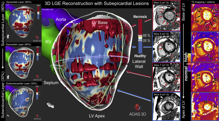
A 64-year-old man presented with severe myocarditis 6 weeks after an initial almost asymptomatic severe acute respiratory syndrome coronavirus-2 (SARS-CoV2) infection. He was found to have a persistent positive swab. Mechanisms explaining myocardial injury in patients with COVID-19 remains unclear, but this case suggests that severe acute myocarditis can develop in the late phase of COVID-19 infection, even after a symptom-free interval.
Un homme de 64 ans a présenté une myocardite grave six semaines après avoir été infecté par le coronavirus du syndrome respiratoire aigu sévère 2 (SRAS-CoV-2), qui n’avait provoqué pratiquement aucun symptôme. Le patient obtenait toujours un résultat positif au test de dépistage du SRAS-CoV-2. On ne connaît pas bien les mécanismes à l’origine des lésions myocardiques observées chez des patients atteints de COVID-19, mais ce cas montre qu’il est possible qu’une myocardite aiguë grave survienne aux stades avancés d’une infection par le SRAS-CoV-2, même après une période sans symptômes.
Copyright © 2020 Canadian Cardiovascular Society. Published by Elsevier Inc. All rights reserved.
Figures
Comment in
-
COVID-19, the heart and returning to physical exercise.Occup Med (Lond). 2020 Oct 27;70(7):467-469. doi: 10.1093/occmed/kqaa154. Occup Med (Lond). 2020. PMID: 32816003 Free PMC article. No abstract available.
References
-
- Biesbroek P.S., Hirsch A., Zweerink A. Additional diagnostic value of CMR to the European Society of Cardiology (ESC) position statement criteria in a large clinical population of patients with suspected myocarditis. Eur Heart J Cardiovasc Imaging. 2018;19:1397–1407. - PubMed
Publication types
MeSH terms
Substances
LinkOut - more resources
Full Text Sources
Miscellaneous
