Review
. 2020 May;11(Suppl 3):S402-S411.
doi: 10.1016/j.jcot.2019.07.006. Epub 2019 Jul 17.

Arthroscopic stabilisation for shoulder instability

Affiliations
Review

Arthroscopic stabilisation for shoulder instability

Konstantinos Fountzoulas et al. J Clin Orthop Trauma. 2020 May.

Abstract

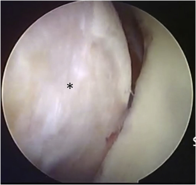
Since its first description over 30 years ago arthroscopic stabilisation has evolved. With improvements in knowledge, surgical techniques and materials technology, arthroscopic bankart repair has become the most widely used method for treating patients with symptomatic anterior shoulder instability. These procedures are typically performed in a younger, high demand patient population after a primary dislocation or to treat recurrent instability. A thorough clinical evaluation is required in the clinic setting not only to fully understand the injury pattern but also consider patient expectations prior to embarking on surgery. Diagnostic imaging will aid the clinician in determining the soft tissue pathology as well as assessing bone loss, which facilitates surgical decision-making. Selected patients may benefit from adjunctive procedures such as a remplissage for an "engaging" Hill-sachs lesion. This review will focus on the indications, pre-operative considerations, surgical techniques and outcomes of arthroscopic stabilisation.

Keywords: Arthroscopic bankart; Arthroscopic stabilisation; Remplissage; Shoulder instability.


Figures

Fig. 1
Fig. 1
Magnetic Resonance arthrogram of the left shoulder showing a classical Bankart lesion.
Fig. 2
Fig. 2
Plain radiograph showing loss of lower part of the sclerotic line representing the anterior glenoid rim (arrows).
Fig. 3
Fig. 3
Computed Tomography (CT) scan of the left shoulder showing glenoid bone loss at the anterior rim.
Fig. 4
Fig. 4
Glenoid track concept. a. 3d CT image showing the relationship between the glenoid rim defect (dotted line) and the Hill-Sachs lesion (arrows) with the arm in maximal abduction and external rotation. b. En face view of the glenoid showing the anterior glenoid bone loss (dotted line shows the margin of the intact glenoid).c. Posterior view of the humerus depicting the track along which the glenoid (dotted margins) would articulate with the humeral head. In this case the margin of the Hill-Sachs lesion (arrows) lies medial to the glenoid track and would indicate that the lesion is likely to engage in the glenoid defect.
Fig. 5
Fig. 5
Arthroscopic demonstration of an engaging Hill-Sachs lesion(*) in a left shoulder seen from the posterior viewing portal.
Fig. 6
Fig. 6
Right shoulder seen through anterosuperior viewing portal. Orange hashed line (r) demonstrates anterior radius whilst blue hashed line represents posterior radius (R). Arrow marks bare spot of the glenoid. Glenoid bone loss is estimated by the formula (R-r) x 100/2R.
Fig. 7
Fig. 7
a. An ALPSA lesion in a right shoulder as seen from the anterosuperior viewing portal. b. Same shoulder after repair – arrows indicate location of anchors.
Fig. 8
Fig. 8
a. Right shoulder – bony Bankart lesion as seen from the anterosuperior viewing portal. b. Same shoulder after repair using the technique described by Sugaya (arrows indicate location of anchors).
Fig. 9
Fig. 9
a. Large (engaging) Hill-Sachs lesion in a left shoulder seen from an anterosuperior viewing portal. The needles mark the sites of anchor placement. b. Appearance after remplissage with the infraspinatus (*) attached to the Hill-Sachs lesion. c. Bursal view showing the sutures tied over the infraspinatus tendon (*).

References

    1. Scheibel M., Nikulka C., Dick A., Schroeder R.J., Popp A.G., Haas N.P. Structural integrity and clinical function of the subscapularis musculotendinous unit after arthroscopic and open shoulder stabilization. Am J Sports Med. 2007;35(7):1153–1161. doi: 10.1177/0363546507299446. - DOI - PubMed
    1. Petrera M., Patella V., Patella S., Theodoropoulos J. A meta-analysis of open versus arthroscopic Bankart repair using suture anchors. Knee Surg Sport Traumatol Arthrosc. 2010;18(12):1742–1747. doi: 10.1007/s00167-010-1093-5. - DOI - PubMed
    1. Carreira D.S., Mazzocca A.D., Oryhon J., Brown F.M., Hayden J.K., Romeo A.A. A prospective outcome evaluation of arthroscopic Bankart repairs: minimum 2-year follow-up. Am J Sports Med. 2006;34(5):771–777. doi: 10.1177/0363546505283259. - DOI - PubMed
    1. Hantes M.E., Venouziou A.I., Liantsis A.K., Dailiana Z.H., Malizos K.N. Arthroscopic repair for chronic anterior shoulder instability: a comparative study between patients with Bankart lesions and patients with combined Bankart and superior labral anterior posterior lesions. Am J Sports Med. 2009;37(6):1093–1098. doi: 10.1177/0363546508331139. - DOI - PubMed
    1. Ricchetti E.T., Ciccotti M.C., O'Brien D.F. Outcomes of arthroscopic repair of panlabral tears of the glenohumeral joint. Am J Sports Med. 2012;40(11):2561–2568. doi: 10.1177/0363546512460834. - DOI - PubMed