. 2019 Jun;4(6):415-428.
doi: 10.1038/s41578-019-0110-7. Epub 2019 May 2.

Material design for lymph node drug delivery

Affiliations

Material design for lymph node drug delivery

Alex Schudel et al. Nat Rev Mater. 2019 Jun.

Abstract

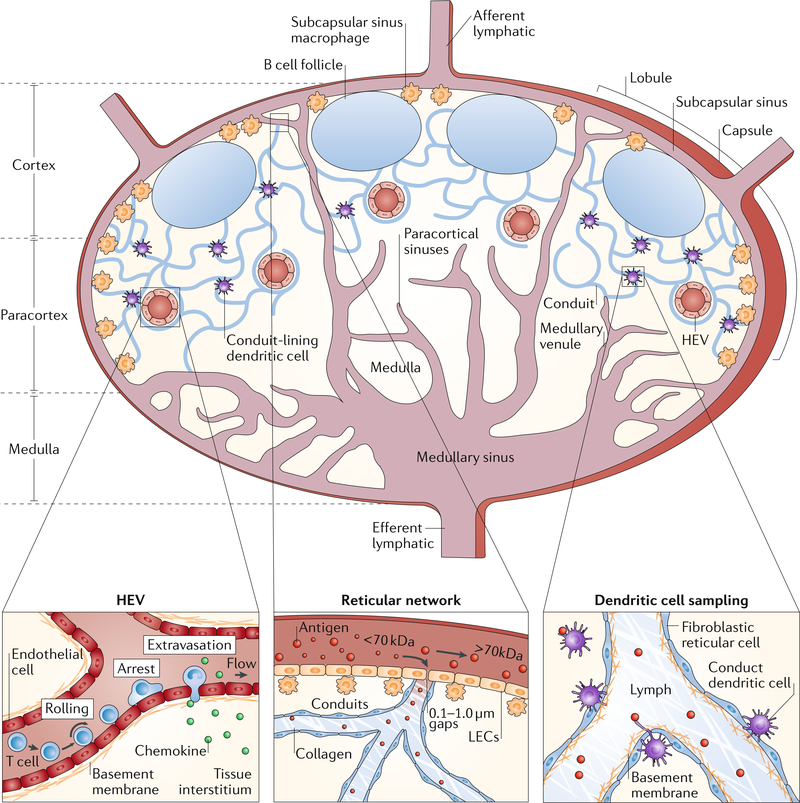
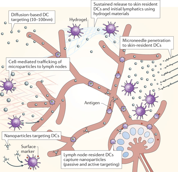
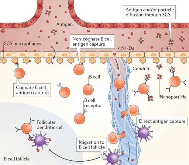
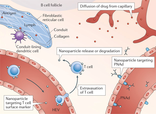
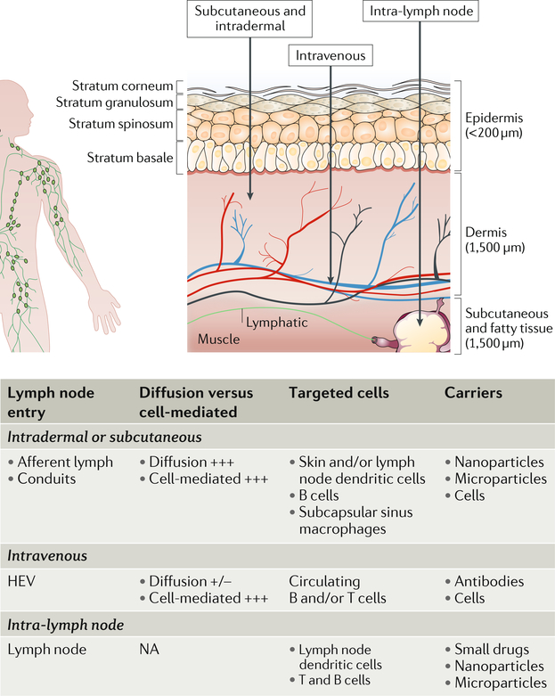
A significant fraction of the total immune cells in the body are located in several hundred lymph nodes, in which lymphocyte accumulation, activation and proliferation are organized. Therefore, targeting lymph nodes provides the possibility to directly deliver drugs to lymphocytes and lymph node-resident cells and thus to modify the adaptive immune response. However, owing to the structure and anatomy of lymph nodes, as well as the distinct localization and migration of the different cell types within the lymph node, it is difficult to access specific cell populations by delivering free drugs. Materials can be used as instructive delivery vehicles to achieve accumulation of drugs in the lymph nodes and to target specific lymph node-resident cell subtypes. In this Review, we describe the compartmental architecture of lymph nodes and the cell and fluid transport mechanisms to and from lymph nodes. We discuss the different entry routes into lymph nodes and how they can be explored for drug delivery, including the lymphatics, blood capillaries, high endothelial venules, cell-mediated pathways, homing of circulating lymphocytes and direct lymph node injection. We examine different nanoscale and microscale materials for the targeting of specific immune cells and highlight their potential for the treatment of immune dysfunction and for cancer immunotherapy. Finally, we give an outlook to the field, exploring how lymph node targeting can be improved by the use of materials.


Conflict of interest statement

Competing interests The authors declare no competing interests.

Figures

Fig. 1 |
Fig. 1 |. Lymph node structure and physiology.
A cross section of a lymph node is shown. The architecture of the lymph node can be divided into distinct areas: fluid-filled lumen structures (lymphatics, high endothelial venules (HEVs), capillaries and sinuses), cellular locations (B cells in follicles, dendritic cells and T cells in the paracortex and macrophages in the subcapsular sinus and medulla) and structural units (cortex, paracortex and medulla). Lymphocyte extravasation occurs in the HEVs. The distribution of antigens within the reticular structure is regulated by haemodynamic size and molecular weight by the capsule and conduit. Circulating lymphocytes enter through the vasculature and exit through the efferent lymphatics. Dendritic cells sample the conduit and conduit structures. LEC, lymphatic endothelial cell. HEV and lymph node cross section adapted from REF, Springer Nature Limited.
Fig. 2 |
Fig. 2 |. Targeting dendritic cells.
Small nanoparticles (10–100 nm in diameter) are taken up by the lymphatics and diffuse to the lymph node to target lymph node-resident dendritic cells (DCs). Large nanoparticles (>100 nm in diameter) and microparticles are mostly entrapped in the interstitial matrix at the site of injection and require capture by peripheral DCs or Langerhans cells (skin) for cell-mediated delivery to lymph nodes. Peripheral and lymph node-resident DCs can be actively targeted using cell subtype-specific surface markers. Hydrogels can be used for the controlled release of molecules in peripheral tissues to enable sustained lymphatic uptake and prolonged DC interactions. Microneedles enable transdermal delivery of particle depots and delivery to DC subtypes that reside within discrete skin layers by adjusting the length of the needles. Lymph node-resident DCs take up passively drained nanoparticles and receive cell-delivered particles.
Fig. 3 |
Fig. 3 |. Targeting B cells.
Subcapsular sinus (SCS) macrophages transfer complementdecorated particles via the complement receptor, whereas they transfer immune complexes bound to particles or materials via Fc receptors to the basal side of the sinus to non-cognate and cognate B cells, respectively. Small antigen can be cleaved from microparticles by proteases and released in the sinus. Antigens then diffuse through the SCS directly to B cells. Materials can enter through gaps (0.1–1.0 μm) in the SCS, enabling diffusion of the materials to B cell follicles for direct B cell sampling. Small materials (<70 kDa) can enter the conduits, where they can be directly captured by B cells. LEC, lymphatic endothelial cell.
Fig. 4 |
Fig. 4 |. Targeting T cells.
Conduit-lining dendritic cells sample antigen for subsequent presentation to proximal T cells. Circulating T cells can be targeted for T cell-mediated nanoparticle trafficking into the lymph node T cell zone. Lymph node blood capillaries that are leaky as a result of disease allow for diffusion-mediated transport to lymph node T cells. Microparticles and nanoparticles can be actively targeted to high endothelial venules (HEVs) using anti-peripheral node addressin (PNAd) antibodies, such as MECA-79, followed by diffusion of the delivered agent into the lymph node.
Fig. 5 |
Fig. 5 |. Route of administration into lymph nodes.
Different regions of skin-draining lymph nodes can be targeted by injections and administration. + and −, scale; HEV, high endothelial venule; NA, not applicable.

References

    1. Sainte-Marie G The lymph node revisited: development, morphology, functioning, and role in triggering primary immune responses. Anat. Rec. 293, 320–337 (2010). - PubMed
    1. Kaldjian EP, Gretz JE, Anderson AO, Shi Y & Shaw S Spatial and molecular organization of lymph node T cell cortex: a labyrinthine cavity bounded by an epithelium-like monolayer of fibroblastic reticular cells anchored to basement membrane-like extracellular matrix. Int. Immunol. 13, 1243–1253 (2001). - PubMed
    1. Chaplin DD Overview of the immune response. J. Allergy Clin. Immunol. 125, S3–S23 (2010). - PMC - PubMed
    1. Murphy K, Travers P, Walport M & Janeway C Janeway’s Immunobiology 8th edn (Garland Science, 2012).
    1. Jeanbart L et al. Enhancing efficacy of anticancer vaccines by targeted delivery to tumor-draining lymph nodes. Cancer Immunol. Res. 2, 436–447 (2014). - PubMed