. 2020 Feb 21;5(2):155-165.
doi: 10.1002/epi4.12382. eCollection 2020 Jun.

Sleep disruption is not observed with brain-responsive neurostimulation for epilepsy

Affiliations

Sleep disruption is not observed with brain-responsive neurostimulation for epilepsy

Leslie Ruoff et al. Epilepsia Open. .

Abstract

Objective: Neurostimulation devices that deliver electrical impulses to the nervous system are widely used to treat seizures in patients with medically refractory epilepsy, but the effects of these therapies on sleep are incompletely understood. Vagus nerve stimulation can contribute to obstructive sleep apnea, and thalamic deep brain stimulation can cause sleep disruption. A device for brain-responsive neurostimulation (RNS® System, NeuroPace, Inc) is well tolerated in clinical trials, but potential effects on sleep are unknown.

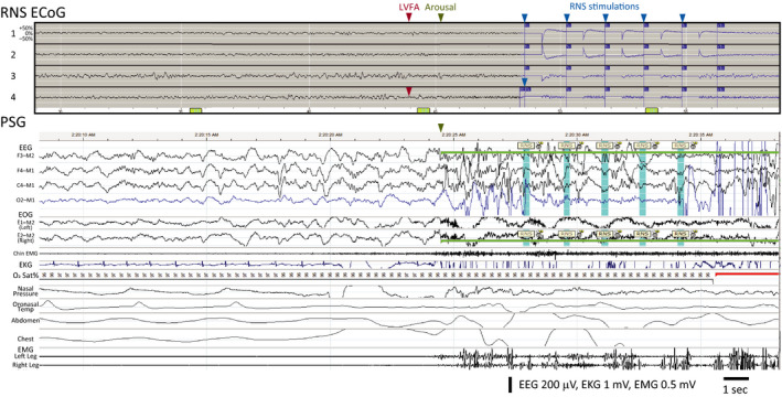
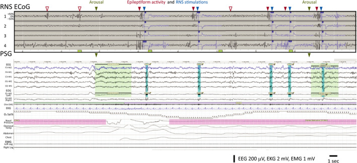
Methods: Six adults with medically refractory focal epilepsy treated for at least six months with the RNS System underwent a single night of polysomnography (PSG). RNS System lead locations included mesial temporal and neocortical targets. Sleep stages and arousals were scored according to standard guidelines. Stimulations delivered by the RNS System in response to detections of epileptiform activity were identified by artifacts on scalp electroencephalography.

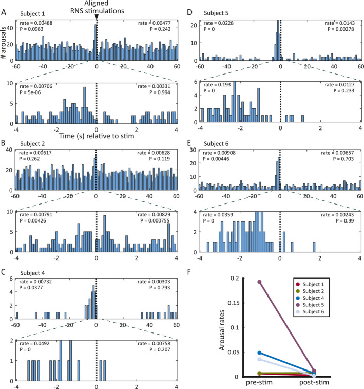
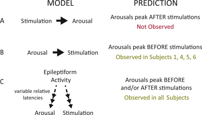
Results: One subject was excluded for technical reasons related to unreliable identification of stimulation artifact on EEG during PSG. In the remaining five subjects, PSG showed fragmented sleep with frequent arousals. Arousal histograms aligned to stimulations revealed a significant peak in arousals just before stimulation. In one of these subjects, the arousal peak began before stimulation and extended ~1 seconds after stimulation. A peak in arousals occurring only after stimulation was not observed.

Significance: In this small cohort of patients, brain-responsive neurostimulation does not appear to disrupt sleep. If confirmed in larger studies, this could represent a potential clinical advantage of brain-responsive neurostimulation over other neurostimulation modalities.

Keywords: RNS; arousal; brain‐responsive neurostimulation; epilepsy; polysomnography; sleep.


Conflict of interest statement

BJ and TKT are employees of, and hold stock options at, NeuroPace, Inc. VRR is a paid consultant for NeuroPace, Inc, but does not have equity or ownership in the company. Other authors declare no relevant conflicts of interest. We confirm that we have read the Journal's position on issues involved in ethical publication and affirm that this report is consistent with those guidelines.

Figures

Figure 1
Figure 1
Temporal relationship between arousals and RNS System stimulations for each subject. A–E, Histograms showing frequency distribution of arousals relative to stimulations aligned at t = 0 for subjects 1 (A), 2, (B), 4 (C), 5 (D), and 6 (E). In each panel, plots show 60‐s (top) and 4‐s (bottom) windows before and after aligned stimulations. Axis labels in A also apply to B–E. All subjects demonstrated a peak in arousals just before stimulation except Subject 2 (B), who showed an increase in arousals both before and after RNS stimulation. Arousal rates shown for each pre‐ and poststimulation window are in units of arousals/RNS stimulation/minute. The P‐values shown in each pre‐ and poststimulation window indicate the probability of observing the actual arousal rate (or higher) by chance given the baseline stimulation and arousal rates for that subject, obtained by randomly shuffling the actual inter‐arousal intervals 100 000 times. F, Plot summarizing the prestimulation (left) and poststimulation (right) arousal rates in the 4‐s windows shown in A‐E. Each bar connects the prestimulation to the poststimulation arousal rate data for one patient. Note that the prestimulation rates tend to be higher than the poststimulation arousal rates (n.s.), which is the opposite of the expected effect if stimulation consistently caused arousals
Figure 2
Figure 2
Epileptiform activity can precede arousal and RNS System stimulation. Data from Subject 4 showing electrocorticogram stored by the RNS System (‘RNS ECoG’) aligned with corresponding polysomnogram (‘PSG’). ECoG Channels 1 and 2 show bipolar recordings from a mesial frontal strip lead, and Channels 3 and 4 show bipolar recordings from a dorsolateral frontal strip lead. PSG shows the subject in stage N3 with an arousal (green highlight) at the end of the epoch and five visually identified stimulation artifacts (blue highlight) labeled “RNS.” The respiratory and leg channels show the absence of any significant respiratory or movement events preceding the arousal. This subject's typical ictal pattern involves attenuation and emergence of low‐voltage fast activity (‘LVFA’) in Channel 4 (red arrowhead), which, here, is followed by arousal from sleep (green arrowhead) and then a sequence of five stimulations (maximum allowed by neurostimulator; blue arrowheads) triggered by detection of this epileptiform pattern (indicated by blue portion of ECoG trace). The temporal sequence of events suggests that epileptiform activity caused both arousal and stimulation
Figure 3
Figure 3
RNS System stimulations can precede and follow arousals. Data from Subject 1, aligned RNS ECoG and PSG as in Figure 1. ECoG Channels 1 and 2 are bipolar recordings from the left hippocampus, and Channels 3 and 4 are recorded from the right hippocampus. ECoG shows frequent bilateral independent epileptiform discharges, some that are undetected by the RNS System (empty red arrowheads) and some that are detected (filled red arrowheads), triggering stimulation (blue arrowheads; note stimulation artifact on top two scalp EEG channels in PSG). PSG captures two arousals (green arrowheads), and stimulations are observed shortly after the first arousal and shortly before the second arousal. The temporal sequence of events suggests that at least some arousals in this subject are independent of stimulations
Figure 4
Figure 4
Models to explain observed results. A, If stimulation causes arousals, then more arousals should follow stimulations than expected by chance, and there should be a higher rate of arousals after than before RNS stimulations. We did not observe either of these outcomes in any patient. B, If arousals trigger brain‐responsive neurostimulation, then arousals should peak before stimulations. This is consistent with observations in all subjects, but it does not entirely explain Subject 2's results, whose arousal peak starts before and extends after stimulation. C, A model in which epileptiform activity causes both arousals and RNS stimulations with variable latencies is consistent with the pre‐ and peristimulation arousal peaks observed in all subjects, including Subject 2

References

    1. Chen Z, Brodie MJ, Liew D, Kwan P. Treatment outcomes in patients with newly diagnosed epilepsy treated with established and new antiepileptic drugs: a 30‐Year Longitudinal Cohort Study. JAMA Neurol. 2018;75:279–86. - PMC - PubMed
    1. Englot DJ, Chang EF. Rates and predictors of seizure freedom in resective epilepsy surgery: an update. Neurosurg Rev. 2014;37:389–404. - PMC - PubMed
    1. Markert MS, Fisher RS. Neuromodulation ‐ science and practice in epilepsy: vagus nerve stimulation, thalamic deep brain stimulation, and responsive neurostimulation. Expert Rev Neurother. 2019;19:17–29. - PubMed
    1. Benbadis SR, Geller E, Ryvlin P, Schachter S, Wheless J, Doyle W, et al. Putting it all together: options for intractable epilepsy: an updated algorithm on the use of epilepsy surgery and neurostimulation. Epilepsy Behav. 2018;88S:33–8. - PubMed
    1. Fisher RS, Velasco AL. Electrical brain stimulation for epilepsy. Nat Rev Neurol. 2014;10:261–70. - PubMed