Review
. 2020 May 18:12:2515841420917781.
doi: 10.1177/2515841420917781. eCollection 2020 Jan-Dec.

Advances in imaging of uveitis

Affiliations
Review

Advances in imaging of uveitis

Alessandro Marchese et al. Ther Adv Ophthalmol. .

Abstract

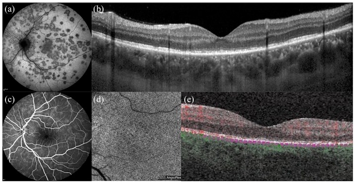
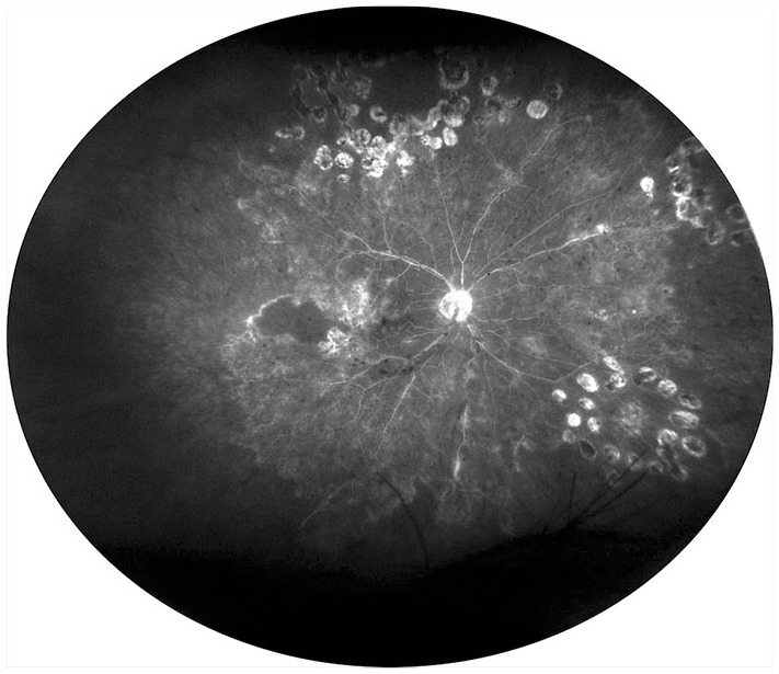
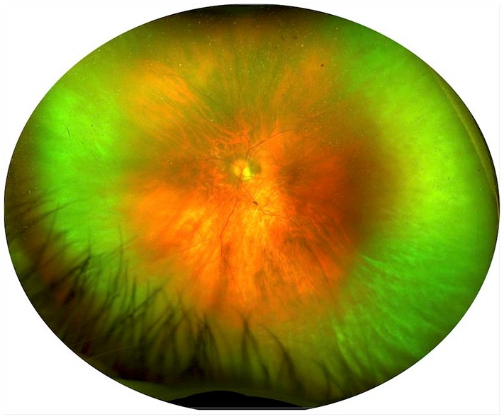
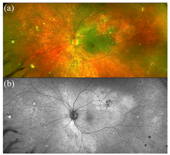
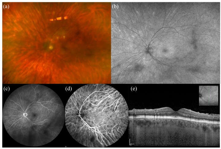
Advances in multimodal imaging have significantly contributed to the management of many uveitis diseases in recent years. The most significant developments include the use of optical coherence tomography to obtain a more accurate and reproducible assessment of ocular inflammation, the application of optical coherence tomography angiography in choroiditis and retinal vasculitis, new possibilities for studying vitritis with ultrawide field imaging, and the most recent applications of fundus autofluorescence in uveitis. In this review, we provide an overview of the most significant advances in multimodal imaging of uveitis achieved in recent years.

Keywords: fundus autofluorescence; optical coherence tomography; optical coherence tomography angiography; ultrawide field imaging; uveitis.


Conflict of interest statement

Conflict of interest statement: AM, AA, AGM, SH, GM, VG and EM have no disclosures. GQ has the following disclosures: Allergan (S), Bayer (S); Novartis (S), Zeiss (S), Allergan (C), Alimera (C), Bausch and Lomb (C), Novartis (C), Bayer (C), Heidelberg (C), and Zeiss (C). F.B. has the following disclosures: Allergan (S), Alimera (S), Bayer (S), Farmila-Thea (S), Schering Pharma (S), Sanofi-Aventis (S), Novagali (S), Pharma (S), Hoffmann-La Roche (S), Genetech (S), and Novartis (S).

Figures

Figure 1.
Figure 1.
Multimodal imaging of multiple evanescent white dot syndrome (MEWDS). (a) Indocyanine green angiography illustrates scattered hypocyanescent dots. (b) OCT shows attenuation and irregularities of the ellipsoid zone. (c) Fluorescein angiography reveals minimal hyper-fluorescence with ‘wreath-like’ lesions. (d and e) On OCT angiography, the choriocapillaris slab shows a normal flow signal.
Figure 2.
Figure 2.
Multimodal imaging of acute posterior multifocal placoid pigment epitheliopathy (APMPPE). In the active phase, (a) fundus photos show multiple yellowish placoid lesions, (b) hypocyanescent on indocyanine green angiography, while (c and d) OCT angiography well delineates the dark areas of choriocapillaris hypoperfusion. Ten days after presentation, (e) fundus autofluorescence shows hyper-autofluorescent lesions.
Figure 3.
Figure 3.
Multimodal imaging of stromal choroiditis. (a) Fundus photograph shows yellowish scattered foci of choroiditis, corresponding to hypocyanescent area on (b) indocyanine green angiography (ICGA). On (c) en-face and (d) cross-sectional B-scan OCT angiography, the areas of choroiditis show the absence of decorrelation signal (black spaces), well correlating with ICGA.
Figure 4.
Figure 4.
Ultrawide field fluorescein angiography of occlusive retinal vasculitis showing extensive areas of capillary nonperfusion, laser scars, and diffused vascular leakage.
Figure 5.
Figure 5.
Ultrawide field imaging of biopsy-proven vitreoretinal lymphoma. The pseudocolor image shows the presence of vitritis along the most peripheral vitreous fibrils with an ‘Aurora Borealis’ appearance, as previously reported.
Figure 6.
Figure 6.
(a) Multimodal imaging of Vogt-Koyanagi-Harada (VKH) syndrome showing mild depigmentation of the retinal pigment epithelium on fundus photograph. (b) Fundus autofluorescence reveals scattered hypo and hyper autofluorescent areas.
Figure 7.
Figure 7.
Multimodal imaging of punctate inner choroidopathy (PIC) complicated by light sensations and paracentral scotomas. (a) Fundus autofluorescence reveals scattered paracentral hyper autofluorescent areas next to a macular hypo autofluorescent scar. These paracentral areas appear hypocyanescent on (b) indocyanine green angiography and mildly hyperfluorescent on (c) fluorescein angiography. (d) OCT illustrates the hyper-reflective macular scar and mild disorganization of outer retinal layers.
Figure 8.
Figure 8.
Multimodal imaging of birdshot chorioretinopathy. (a) Fundus photograph shows yellowish scattered foci of choroiditis, corresponding to hyper autofluorescent lesions on (b) fundus autofluorescence. (c) Fluorescein angiography shows some hypo fluorescent areas corresponding to vitreous opacities and (d) indocyanine green angiography reveals the presence of hypocyanescent foci of choroiditis. (e) OCT does not show significant changes in the macular scan.
Figure 9.
Figure 9.
Multimodal imaging of acute zonal occult outer retinopathy (AZOOR). (a) Fundus photography shows minimal outer retina and retinal pigment epithelium changes, while (b) FAF depicts very well the typical trizonal pattern. In particular, normal autofluorescence is observed outside a demarcating line (zone 1), speckled hyper-autofluorescence is seen within the active lesion (zone 2), and hypo-autofluorescence is detected where chorioretinal atrophy is developed (zone 3). Panels on the bottom row show (c) ICGA in the late phase, (d) infrared fundus image and (e) OCT.
Figure 10.
Figure 10.
Fundus autofluorescence of serpiginous choroiditis showing the annual progression of the disease in the areas of activity (arrows). The active borders of choroiditis appear hyper-autofluorescent and the inactive areas show hypo-autofluorescence.

References

    1. Giuffre C, Miserocchi E, Modorati G, et al. Central serous chorioretinopathylike mimicking multifocal vitelliform macular dystrophy: an ocular side effect of mitogen/extracellular signal-regulated kinase inhibitors. Retin Cases Brief Rep 2018; 12: 172–176. DOI: 10.1097/ICB.0000000000000491. - DOI - PubMed
    1. Marchese A, Romano F, Cicinelli MV, et al. Chorioretinal punched-out lesions in pseudoxanthoma elasticum. Retina 2018; 38: e43–e44. DOI: 10.1097/IAE.0000000000002052. - DOI - PubMed
    1. Corbelli E, Miserocchi E, Marchese A, et al. Ocular toxicity of mirvetuximab. Cornea 2019; 38: 229–232. DOI: 10.1097/ICO.0000000000001805. - DOI - PubMed
    1. Giuffre C, Miserocchi E, Marchese A, et al. Widefield OCT angiography and ultra-widefield multimodal imaging of Susac syndrome. Eur J Ophthalmol. Epub ahead of print 10 April 2019. DOI: 10.1177/1120672119843281. - DOI - PubMed
    1. Cicinelli MV, Marchese A, Miserocchi E, et al. Retinal and choroidal changes of vitreoretinal lymphoma from active to remission phase after intravitreal rituximab. Ocul Immunol Inflamm. Epub ahead of print 8 August 2019. DOI: 10.1080/09273948.2019.1616769. - DOI - PubMed

LinkOut - more resources