. 2022 Apr 19;225(8):1435-1446.
doi: 10.1093/infdis/jiaa329.

Screening of a Library of Recombinant Schistosoma mansoni Proteins With Sera From Murine and Human Controlled Infections Identifies Early Serological Markers

Affiliations

Screening of a Library of Recombinant Schistosoma mansoni Proteins With Sera From Murine and Human Controlled Infections Identifies Early Serological Markers

Cécile Crosnier et al. J Infect Dis. .

Abstract

Background: Schistosomiasis is a major global health problem caused by blood-dwelling parasitic worms, which is currently tackled primarily by mass administration of the drug praziquantel. Appropriate drug treatment strategies are informed by diagnostics that establish the prevalence and intensity of infection, which, in regions of low transmission, should be highly sensitive.

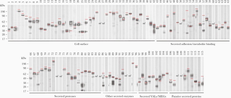
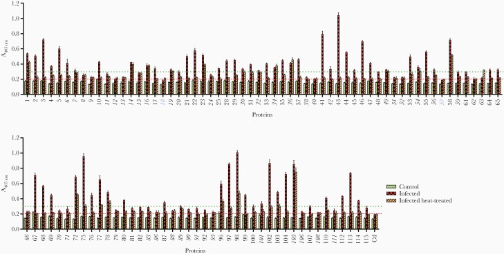
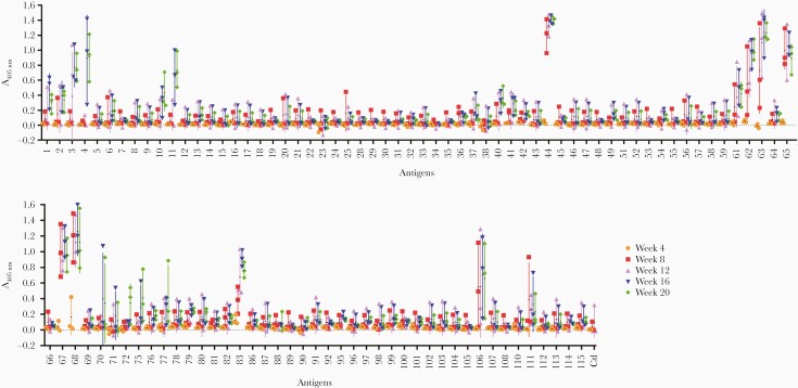
Methods: To identify sensitive new serological markers of Schistosoma mansoni infections, we have compiled a recombinant protein library of parasite cell-surface and secreted proteins expressed in mammalian cells.

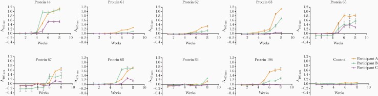
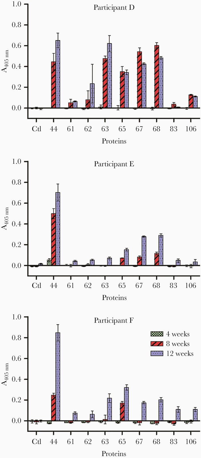
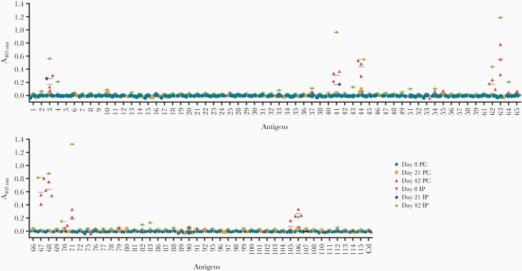
Results: Together with a time series of sera samples from volunteers experimentally infected with a defined number of male parasites, we probed this protein library to identify several markers that can detect primary infections with as low as 10 parasites and as early as 5 weeks postinfection.

Conclusions: These new markers could be further explored as valuable tools to detect ongoing and previous S mansoni infections, including in endemic regions where transmission is low.

Keywords: Schistosoma mansoni; antibodies; schistosomiasis; serology.


Figures

Figure 1.
Figure 1.
A library of 115 recombinant cell-surface and secreted proteins from Schistosoma mansoni expressed as secreted enzymatically monobiotinylated recombinant proteins in HEK293 cells. Supernatants were resolved by sodium dodecyl sulfate polyacrylamide gel electrophoresis (SDS-PAGE) under reducing conditions, blotted, and detected with streptavidin-conjugated horseradish peroxidase. Approximately one third of the protein library consists of membrane-tethered surface proteins, whereas the remainder of the library corresponds to secreted proteins as indicated. Their predicted molecular mass is indicated by a red line. Three proteins (52, 57, 86) migrated faster than expected; 6 proteins (42, 68, 80, 89, 91, 102) exhibited evidence of partial processing; 13 proteins were not detected by Western blotting.
Figure 2.
Figure 2.
The majority of recombinant proteins are immunoreactive to sera from individuals living in schistosomiasis-endemic areas and contain heat-labile conformational epitopes. Recombinant proteins were probed with pooled sera from individuals living in schistosomiasis-endemic areas (“infected,” red checked bars) or individuals from the United Kingdom who have never been infected (“control,” green dotted bars). To test for the presence of heat-labile epitopes, recombinant proteins were also heat-treated (80°C, 10 minutes) before being exposed to immune sera (“infected heat treated,” orange hatched bars). All except 2 proteins (18 and 57, shown in blue) were seropositive, as determined by Aprotein > Acontrol + 3SDcontrol ( = 0.201) (red dashed line), where control is the rat Cd4d3 + 4 protein tag. High immunoreactivity was determined as Aprotein > 0.3 (green dotted line). Proteins that exhibited little or no loss of reactivity after heat treatment are shown in italics, including 16 highly reactive proteins (6, 7, 14, 16, 19, 30, 32, 34, 36, 42, 49, 54, 56, 89, 101, 105). All measurements were performed in triplicate; error bars = standard deviation.
Figure 3.
Figure 3.
Identification of early markers of infection in human volunteers experimentally infected with Schistosoma mansoni male cercariae. Three individuals were each infected with 30 male cercariae, and their antibody response to 103 Schistosoma antigens were monitored every 4 weeks over a period of 20 weeks. The number of positive antigens increased over time with the most highly immunoreactive antigens containing saposin domains (proteins 44, 61, 62, 63, and 65) or belonging to the cathepsin family of proteases (proteins 67, 68, and 83). Colored symbols represent time point readings for each individual. Data points represent mean ± standard deviation; n = 3.
Figure 4.
Figure 4.
Kinetics of human antibody response to early markers of infection using sera from experimental infections by Schistosoma mansoni. Immunoreactivity to S mansoni antigens were quantified on a weekly basis in 3 individuals infected with 30 male cercariae. Reactivity to proteins 44 and 65 could be detected in all volunteers as early as 5 weeks postinfection. The control antigen corresponds to the rat Cd4d3 + 4 protein tag. Data points represent mean ± standard deviation; n = 3.
Figure 5.
Figure 5.
Early reactivity to Schistosoma mansoni antigens is detectable in individuals challenged with just 10 cercariae. The immune response from 3 human volunteers infected with 10 male S mansoni cercariae was monitored at 4, 8, and 12 weeks postinfection. Reactivity to antigens 44 and 65 was detected in all participants at 8 weeks and as early as 4 weeks in the case of participant E and antigen 44. With the exception of protein 83, all volunteers were immunoreactive for the antigens tested at 12 weeks postinfection. Data points represent mean ± standard deviation; n = 3.
Figure 6.
Figure 6.
Analysis of the acquired antibody response to Schistosoma mansoni antigens in experimentally infected mice. Individual sera from 3 mice infected percutaneously (PC) or pooled sera from 3 mice infected intraperitoneally (IP) were analyzed at 8, 21, and 42 days postinfection. The aim was to try and capture the antibody reactivity at different stages of parasite maturation: schistosomule at 8 days, immature adult at 21 days, and mature adult at 42 days. Proteins 44 and 3 were immunopositive at day 21, and 8 additional proteins (10, 41, 62, 63, 64, 67, 68, 106) were immunopositive at day 42. Each data point represents the average of triplicate experiments; error bars = standard deviation.

References

    1. World Health Organization. Schistosomiasis and soil-transmitted helminthiases: number of people treated in 2016. Wkly Epidemiol Rec 2017; 92:749–60. - PubMed
    1. Colley DG, Bustinduy AL, Secor WE, King CH. Human schistosomiasis. Lancet 2014; 383:2253–64. - PMC - PubMed
    1. Cousin CE, Stirewalt MA, Dorsey CH. Schistosoma mansoni: ultrastructure of early transformation of skin- and shear-pressure-derived schistosomules. Exp Parasitol 1981; 51:341–65. - PubMed
    1. Cheever AW, Macedonia JG, Mosimann JE, Cheever EA. Kinetics of egg production and egg excretion by Schistosoma mansoni and S. japonicum in mice infected with a single pair of worms. Am J Trop Med Hyg 1994; 50:281–95. - PubMed
    1. Burke ML, Jones MK, Gobert GN, Li YS, Ellis MK, McManus DP. Immunopathogenesis of human schistosomiasis. Parasite Immunol 2009; 31:163–76. - PubMed

Publication types