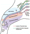
Detection of Neutrophils in the Sciatic Nerve Following Peripheral Nerve Injury
- PMID: 32524483
- PMCID: PMC11131227
- DOI: 10.1007/978-1-0716-0585-1_16
Detection of Neutrophils in the Sciatic Nerve Following Peripheral Nerve Injury
Abstract
Injury to the sciatic nerve leads to degeneration and debris clearance in the area distal to the injury site, a process known as Wallerian degeneration. Immune cell infiltration into the distal sciatic nerve plays a major role in the degenerative process and subsequent regeneration of the injured motor and sensory axons. While macrophages have been implicated as the major phagocytic immune cell participating in Wallerian degeneration, recent work has found that neutrophils, a class of short-lived, fast responding white blood cells, also significantly contribute to the clearance of axonal and myelin debris. Detection of specific myeloid subtypes can be difficult as many cell-surface markers are often expressed on both neutrophils and monocytes/macrophages. Here we describe two methods for detecting neutrophils in the axotomized sciatic nerve of mice using immunohistochemistry and flow cytometry. For immunohistochemistry on fixed frozen tissue sections, myeloperoxidase and DAPI are used to specifically label neutrophils while a combination of Ly6G and CD11b are used to assess the neutrophil population of unfixed sciatic nerves using flow cytometry.
Keywords: Flow cytometry; Immunohistochemistry; Neutrophil; Peripheral nerve injury; Sciatic nerve; Wallerian degeneration.
Figures


References
-
- Waller AV (1850) Experiments on the section of glossopharyngeal and hypoglossal nerves of the frog and observations of the alternatives produced thereby in the structure of their primitive fibres. Philos Trans R Soc Lond B Biol Sci 140:423–429
-
- Vargas ME, Barres BA (2007) Why is Wallerian degeneration in the CNS so slow? Annu Rev Neurosci 30:153–179 - PubMed
-
- Stoll G, Griffin JW, Li CY, Trapp BD (1989) Wallerian degeneration in the peripheral nervous system: participation of both Schwann cells and macrophages in myelin degradation. J Neurocytol 18:671–683 - PubMed
Publication types
MeSH terms
Substances
Grants and funding
LinkOut - more resources
Full Text Sources
Medical
Research Materials

