Review
. 2020 Jul;21(7):793-811.
doi: 10.3348/kjr.2019.0822.

Normal and Abnormal Postoperative Imaging Findings after Gastric Oncologic and Bariatric Surgery

Affiliations
Review

Normal and Abnormal Postoperative Imaging Findings after Gastric Oncologic and Bariatric Surgery

Cheong Il Shin et al. Korean J Radiol. 2020 Jul.

Abstract

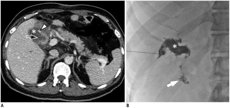
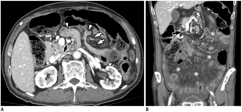
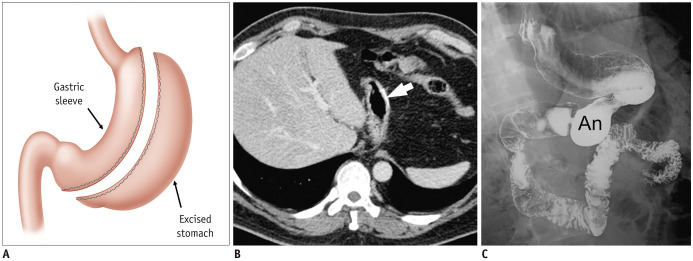
Surgical resection remains the primary choice of treatment and the only potentially curative option for gastric carcinoma, and is increasingly performed laparoscopically. Gastric resection represents a challenging procedure, with a significant morbidity and non-negligible postoperative mortality. The interpretation of imaging after gastric surgery can be challenging due to significant modifications of the normal anatomy. After the surgery, the familiarity with expected imaging appearances is crucial for diagnosis and appropriate management of potentially life-threatening complications in patients who underwent gastric surgery. We review various surgical techniques used in gastric surgery and describe fluoroscopic and cross-sectional imaging appearances of normal postoperative anatomic changes as well as early and late complications after gastric surgery.

Keywords: CT; Postoperative complications; Stomach; Stomach cancer; Surgical techniques.


Conflict of interest statement

The authors have no potential conflicts of interest to disclose.

Figures

Fig. 1
Fig. 1. Postoperative anatomy in subtotal distal gastrectomy with Billroth I anastomosis.
A. Illustration shows gastroduodenostomy in end-to-end fashion. B, C. On axial CT scans, high attenuating surgical materials (arrowheads) are seen at anastomotic site between remnant stomach (S) and duodenum (D), and along lesser curvature suture site. Note that gastric fundus (F) and esophagogastric junction (arrow) are intact.
Fig. 2
Fig. 2. Postoperative anatomy in subtotal distal gastrectomy with Billroth II anastomosis.
Illustration (A) and serial axial CT images (B–D) show loop gastrojejunostomy in antecolic and ipsiperistalic manner. On axial CT images, jejunal segment attached at left side of remnant stomach (S) is traced into duodenum (Du), indicating afferent loop (A), then other jejunal segment attached at right side of remnant stomach goes into efferent loop (E). Peristaltic direction of remnant stomach and efferent loop is same, so-called ipsiperistaltic. In addition, afferent and efferent loops are located anterior to transverse colon (Co), indicating antecolic anastomosis. Duodenal stump (Du) is easily identified with surgical materials (arrowhead) in right subhepatic space.
Fig. 3
Fig. 3. Postoperative anatomy in total gastrectomy with Roux-en-Y reconstruction.
Illustration (A) and serial axial CT images (B–E) show Roux-en-Y esophagojejunostomy after total gastrectomy. CT images show multiple surgical clips at esophagojejunostomy site (black arrowheads), jejunal stump (white arrow), jejunojejunostomy site (black arrows), and duodenal stump (white arrowhead). Alimentary limb (A) is located anterior to transverse colon (Co), indicating antecolic anastomosis. Bi = biliopancreatic limb
Fig. 4
Fig. 4. Postoperative anatomy in pylorus-preserving gastrectomy.
A. Illustration shows gastrogastrostomy after pylorus-preserving gastrectomy. Note that hepatic (open arrow) and pyloric (open arrowhead) branches of vagus nerve were saved. An = antrum, Bo = gastric upper body, Du = duodenum. B. On axial CT image, pyloric canal (arrowhead) and normal triangular duodenal bulb (Du) are intact. Contour deformity at gastrogastric anastomosis (arrows) between prepyloric antrum (An) and gastric upper body (Bo) is also noted. Compared to distal gastrectomy, visualization of remnant stomach including fundus, is same, but intact pylorus is a different point.
Fig. 5
Fig. 5. Postoperative anatomy in proximal gastrectomy with double tract reconstruction.
Illustration (A) and contrast swallow study (B) show double tract reconstruction after proximal gastrectomy composed of Roux-en-Y esophagojejunostomy (arrowhead) and end-to-side gastrojejunostomy between remnant stomach (S) and proximal efferent jejunal loop (E), allowing food passage flow simultaneously to remnant stomach (blue arrow) and jejunum (red arrow). A = afferent jejunal loop, D = duodenum. Coronal CT image (C) also well demonstrates remnant stomach (S) and continued duodenal loop (D) as well as efferent jejunal loop (E).
Fig. 6
Fig. 6. Anastomotic leakage after Billroth I reconstruction.
63-year-old male patient underwent distal gastrectomy with Billroth I reconstruction for advanced gastric cancer. A. On CT, which was taken at postoperative day 3 for fever evaluation, air-containing fluid collections (*) are noted anterior to pancreas. B. Contrast swallow study was performed on postoperative day 11 showing patent gastroduodenal anastomosis but extraluminal leakage (arrows). Opacification at percutaneous drainage tube is also noted (arrowheads).
Fig. 7
Fig. 7. Anastomotic leakage after total gastrectomy and Roux-en-Y reconstruction.
72-year-old male patient underwent total gastrectomy with Roux-en-Y reconstruction. A. Contrast swallow study, which was performed 5 days after surgery, shows contrast leakage (black arrow) at left side of esophagojejunostomy (EJstomy). Short and blind jejunal stump (arrowheads) appears as irregular contrast-filling structure below esophagojejunostomy (EJstomy), which can be mistaken for leak. B. On noncontrast CT taken immediately after contrast swallow study, small pooling of contrast material (white arrow) is seen outside surgical materials of esophagojejunostomy, indicating leakage. Small pleural effusion (*) in left lower thorax is also noted.
Fig. 8
Fig. 8. Duodenal stump leakage.
60-year-old male patient underwent subtotal gastrectomy with Billroth II anastomosis. A. On CT for postoperative fever evaluation, small fluid collection (*) is noted around duodenal stump with defect (open arrow) at lateral wall. Tiny high attenuating structure abutting on duodenal stump is surgical material (arrowhead). B. Cavitogram during percutaneous drainage well demonstrates complicated fluid collection (*) communicating with duodenum (arrow).
Fig. 9
Fig. 9. Gastric stasis after pylorus-preserving gastrectomy.
A. Plain abdominal radiograph on postoperative day 6 shows distended remnant stomach (S) filled with residual food materials. B. On contrast swallow study, which was taken on postoperative day 7, pyloric canal (arrow) is severely narrowed without relaxation, suggesting impaired pyloric function. Note that gastrogastric anastomosis (arrowheads) is widely patent.
Fig. 10
Fig. 10. Acute pancreatitis after total gastrectomy.
52-year-old male patient underwent total gastrectomy with splenectomy for advanced gastric cancer (pT4aN1) at gastric cardia. Two days after surgery, amount of drained fluid increased, and amylase level in drain was increased to 2076 U/L. A. Postoperative axial CT at day 4 shows decreased enhancement at pancreas tail and surrounding peripancreatic fat infiltration (arrowheads), suggesting acute pancreatitis. B. Coronal CT image at postoperative day 14 shows interval development of complicated fluid collections around esophagojejunostomy (arrows) and perihepatic space (*). Therefore, leakage at esophagojejunostomy site (open arrow) is strongly suspected, although wall defect is not delineated on CT.
Fig. 11
Fig. 11. Omental infarction.
A. On axial CT taken at 14 days after laparoscopy-assisted distal gastrectomy, small fluid collection and fat stranding are noted in omentum (arrows). B. On follow-up CT after 1 year, smaller, well-defined, heterogeneously attenuated mass with high attenuating rim (arrowhead) is delineated in same area. Note that fat attenuation of lesion totally disappears.
Fig. 12
Fig. 12. Ischemia of left lobe of liver after gastric surgery.
45-year-old male patient underwent open total gastrectomy for advanced gastric cancer. A. Maximum intensity projection image of preoperative CT well demonstrates replaced left hepatic artery (arrows) arising from left gastric artery (arrowhead). During operation, left gastric artery was ligated at proximal portion. Then, levels of serum aspartate aminotransferase and alanine aminotransferase increased over 1000 IU/L at postoperative day 1. B. On postoperative day 3 CT, enhancement in left lateral segment of liver becomes markedly decreased (*), suggesting ischemia. C. On follow-up CT after 3 months, perfusion to left lateral hepatic segment restores to normal range with mild parenchymal volume decrease (open arrows).
Fig. 13
Fig. 13. Remnant gastric infarct.
77-year-old male patient with long history of hypertension and diabetes underwent laparoscopy-assisted distal gastrectomy with Billroth II reconstruction for advanced gastric cancer. On axial (A) and coronal (B) CT images obtained at postoperative day 5 for postoperative fever evaluation, lesser curvature and anterior wall of remnant stomach are poorly enhanced (black arrows) compared to normal enhancement of gastric fundus (white arrow). Total gastrectomy was then performed, and transmural infarct was confirmed at lesser curvature to anterior wall of remnant gastric specimen (not shown).
Fig. 14
Fig. 14. Afferent loop syndrome due to adhesive ileus after subtotal distal gastrectomy with Billroth II reconstruction.
56-year-old male patient underwent subtotal gastrectomy with Billroth II reconstruction for early gastric cancer 2 years ago. He presented with abdominal pain and nausea. A, B. On axial CT images, afferent limb (*) is markedly dilated with fluid, while efferent limb is not. B. Transition zone (arrow) is noted immediately distal to Treitz ligament in left abdomen. It shows beak-like appearance suggesting adhesive ileus. S = remnant stomach
Fig. 15
Fig. 15. Internal hernia after total gastrectomy.
A. Axial CT image obtained at postoperative 1-year shows protrusion of small bowel (arrowheads) into anterior abdominal cavity passing between superior mesenteric artery (black arrow) and distal mesenteric arterial branches (white arrows), so-called mushroom sign. B. On coronal CT image, decreased caliber of superior mesenteric vein with beaked appearance (arrow), and extensive mesenteric congestion (*) are also seen. Laparotomy reveals Petersen's hernia.
Fig. 16
Fig. 16. Adjustable gastric banding and postoperative images.
Illustration (A) and CT scanogram (B) show expected appearance after adjustable gastric banding. Gastric band (black arrow) overlies left side of spine just below level of left hemidiaphragm. Angle formed by longitudinal axis of gastric band and spine is called phi (φ) angle. It is usually between 4° and 58° in normal conditions. Reservoir port (arrowhead) overlies anterior abdominal wall. Connecting tube location can vary within peritoneum. C. On axial CT image, inflatable balloon cuff of band (white arrows) is positioned around proximal stomach.
Fig. 17
Fig. 17. Gastric band erosion.
A. Coronal CT image shows lateral part of gastric band (black arrow) has eroded into gastric lumen (S). B. On axial CT image, liver abscess (*), and peritoneal infiltration (arrowhead) suggesting peritonitis are seen around tube (white arrow).
Fig. 18
Fig. 18. Sleeve gastrectomy.
A. Illustration shows normal surgical anatomy after sleeve gastrectomy. Stomach is resected along greater curvature of fundus, body, and proximal antrum, producing narrow, banana-shaped gastric sleeve along lesser curvature. B. On axial CT image, metallic suture (arrow) is observed in new greater curvature of stomach. C. Contrast swallow study shows decrease in gastric volume with tubular morphology. Note that distal antrum (An) is intact.
Fig. 19
Fig. 19. Roux-en-Y gastric bypass.
A. Illustration of expected postsurgical anatomy following Roux-en-Y gastric bypass shows gastric pouch, excluded gastric remnant, antegastric Roux limb, jejunal stump, and biliopancreatic limb. B, C. Axial CT images delineate gastric pouch (GP), excluded stomach (ES), Roux limb (RL), and its jejunal stump (JS).

References

    1. Soerjomataram I, Lortet-Tieulent J, Parkin DM, Ferlay J, Mathers C, Forman D, et al. Global burden of cancer in 2008: a systematic analysis of disability-adjusted life-years in 12 world regions. Lancet. 2012;380:1840–1850. - PubMed
    1. Torre LA, Bray F, Siegel RL, Ferlay J, Lortet-Tieulent J, Jemal A. Global cancer statistics, 2012. CA Cancer J Clin. 2015;65:87–108. - PubMed
    1. Lee DH, Kim SH, Joo I, Hur BY, Han JK. Comparison between 18F-FDG PET/MRI and MDCT for the assessment of preoperative staging and resectability of gastric cancer. Eur J Radiol. 2016;85:1085–1091. - PubMed
    1. Inokuchi M, Otsuki S, Fujimori Y, Sato Y, Nakagawa M, Kojima K. Systematic review of anastomotic complications of esophagojejunostomy after laparoscopic total gastrectomy. World J Gastroenterol. 2015;21:9656–9665. - PMC - PubMed
    1. Xiong JJ, Altaf K, Javed MA, Nunes QM, Huang W, Mai G, et al. Roux-en-Y versus Billroth I reconstruction after distal gastrectomy for gastric cancer: a meta-analysis. World J Gastroenterol. 2013;19:1124–1134. - PMC - PubMed

Publication types

MeSH terms