. 2020 Oct;14(10):2646-2659.
doi: 10.1002/1878-0261.12745. Epub 2020 Jul 13.

Serum small extracellular vesicle-derived LINC00853 as a novel diagnostic marker for early hepatocellular carcinoma

Affiliations

Serum small extracellular vesicle-derived LINC00853 as a novel diagnostic marker for early hepatocellular carcinoma

Soon Sun Kim et al. Mol Oncol. 2020 Oct.

Abstract

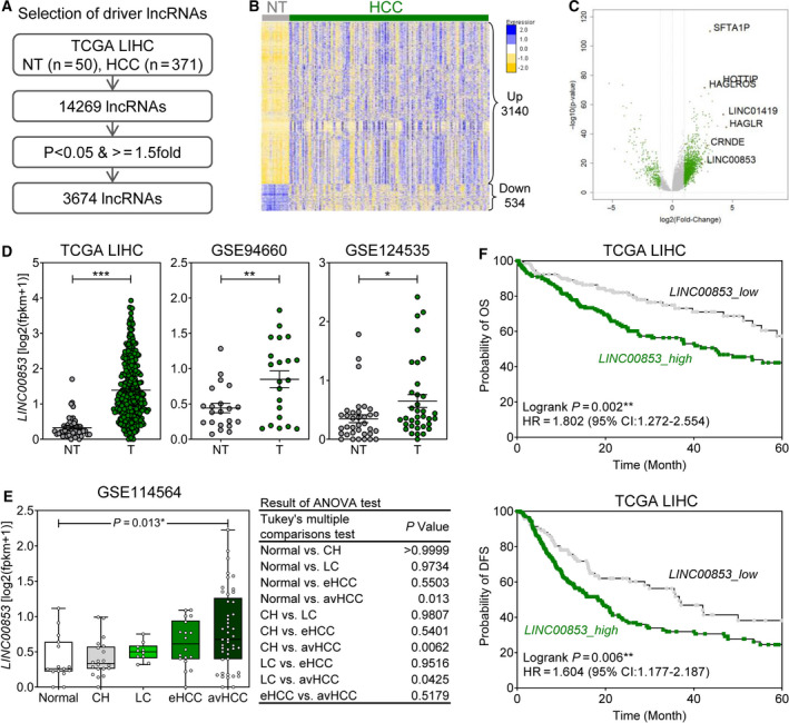
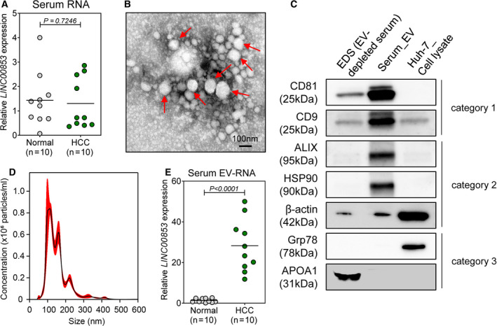
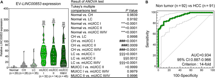
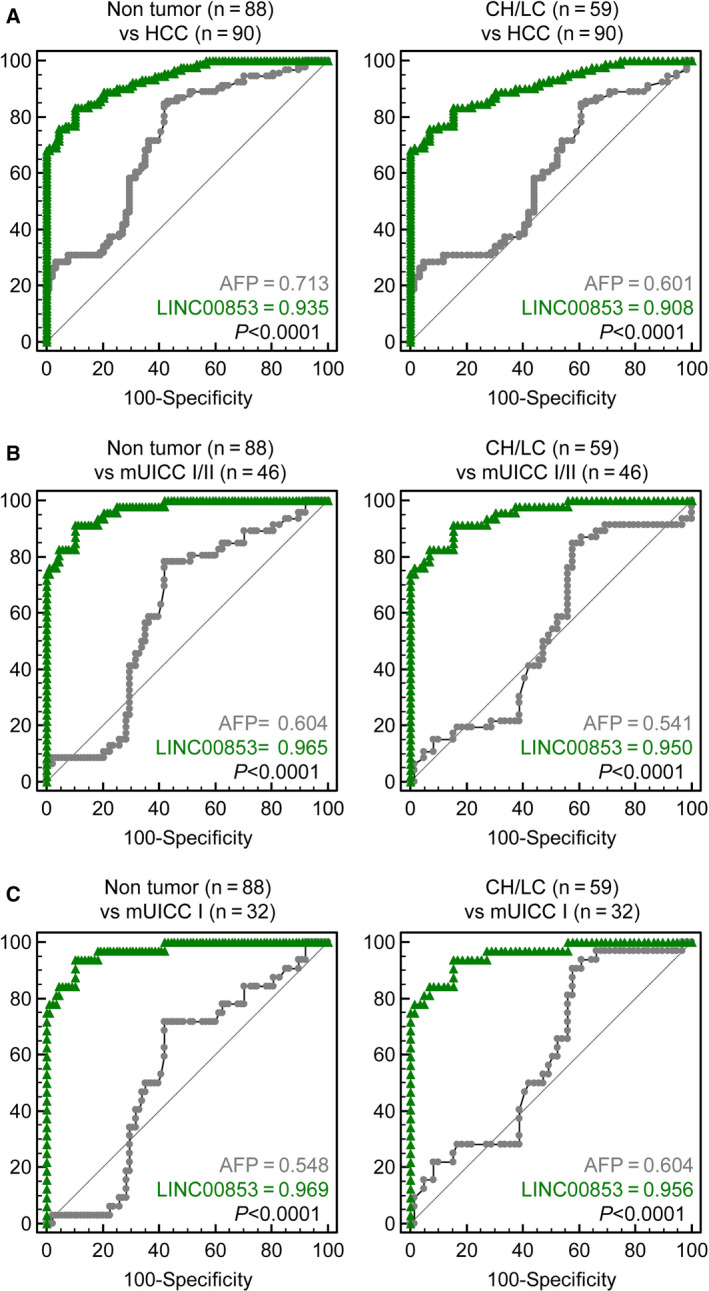
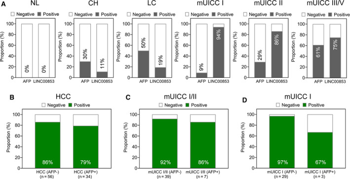
This study aimed to identify novel long noncoding RNA (lncRNA) biomarkers for hepatocellular carcinoma (HCC) using publicly available tissue genomic datasets and validate their diagnostic utility for early-stage HCC. Differentially expressed lncRNAs between 371 HCC and 50 nontumor tissues were obtained from The Cancer Genome Atlas liver hepatocellular carcinoma (TCGA_LIHC) project. Subsequently, the expression of the serum- and extracellular vesicle (EV)-derived lncRNA was assessed in 10 patients with HCC and 10 healthy controls using RT-qPCR. The candidate lncRNAs were validated in 90 HCC and 92 non-HCC (29 healthy control, 28 chronic hepatitis, 35 liver cirrhosis) patients. The sensitivity, specificity, and area under the receiver operating characteristic curve (AUC) were calculated for the candidate lncRNAs and the current HCC biomarker, alpha-fetoprotein (AFP). SFTA1P, HOTTIP, HAGLROS, LINC01419, HAGLR, CRNDE, and LINC00853 were markedly upregulated in HCC in TCGA_LIHC dataset. Among them, LINC00853 has not been reported in relation to HCC before. In patients with HCC, only expression of small EV-derived LINC00853 (EV-LINC00853) was increased. EV-LINC00853 showed excellent discriminatory ability in the diagnosis of all-stage HCC (AUC = 0.934, 95% confidence interval = 0.887-0.966). Moreover, using a 14-fold increase and 20 ng·mL-1 as cutoffs for EV-LINC00853 expression and AFP level, respectively, EV-LINC00853 was found to have a sensitivity of 93.75% and specificity of 89.77%, while AFP showed only 9.38% sensitivity and 72.73% specificity for the diagnosis of early-stage HCC (mUICC stage I). EV-LINC00853 had a positivity of 97% and 67% in AFP-negative and AFP-positive early HCC, respectively. Serum EV-derived LINC00853 may be a novel potential diagnostic biomarker for early HCC, especially for AFP-negative HCC.

Keywords: LINC00853; biomarker; extracellular vesicles; hepatocellular carcinoma; long noncoding RNA.


Conflict of interest statement

The authors declare no conflict of interest.

Figures

Fig. 1
Fig. 1
Correlation between clinical findings and LINC00853 overexpression in HCC cohorts. (A) The strategy to identify novel lncRNA markers for HCC. (B) Heatmap of 3674 HCC‐associated lncRNA signatures in the TCGA_LIHC dataset. (C) Volcano plot representation of differentially expressed lncRNA signatures in the nontumor and the HCC cohorts. (D) LINC00853 expression in the nontumor and the HCC cohorts in three HCC RNA‐Seq datasets (TCGA_LIHC, GSE94660, and GSE124535). Welch's t‐test; *P < 0.05, **P < 0.01, ***P < 0.001. (E) Changes in expression of 10 candidate marker genes in patients with different types and severity of liver disease in GSE114564 dataset. Statistically significant differences were determined using one‐way ANOVA with Tukey's multiple comparisons test. (F) The Kaplan–Meier survival curves for overall survival (left) and disease‐free survival (right) based on LINC00853 expression in patients with HCC in TCGA_LIHC dataset.
Fig. 2
Fig. 2
Expression of LINC00853 in patient serum samples and serum‐derived EVs. (A) Scatter plot of LINC00853 expression in the sera of healthy subjects (n = 10) and patients with HCC (n = 10). Statistically significant differences were determined using Welch's t‐test. Black horizontal lines denote means. (B) TEM image showing the spherical morphology of the isolated small EVs (diameter = ~ 100 nm), bar = 100 nm. (C) Representative immunoblots showing the expression of EV markers in the isolated EVs, according to MISEV 2018 guidelines. EDS and Huh‐7 cell lysate were used as controls. (D) The concentration and size distribution of EVs in the serum of a patient with HCC, as determined by NTA. (E) Scatter plot of LINC00853 expression in serum‐derived EVs of healthy subjects (n = 10) and patients with HCC (n = 10). Statistically significant differences were determined using Welch's t‐test. Black horizontal lines denote sample means. Target gene expression was calculated relative to that of HMBS.
Fig. 3
Fig. 3
Expression of EV‐LINC00853 and its diagnostic performance in the validation cohort. (A) Violin plot of EV‐LINC00853 expression, as measured by RT–qPCR. Statistically significant differences were determined using the one‐way ANOVA with Tukey's multiple comparisons test. Black horizontal lines denote means, and error bars represent SEM. Compared to healthy liver; *P < 0.05, **P < 0.01, ***P < 0.001, compared to CH; #P < 0.05, ##P < 0.01, ###P < 0.001, compared to LC; §P < 0.05, §§P < 0.01, §§§P < 0.001. (B) Analysis of EV‐LINC00853 ROC curve in patients with HCC vs control (healthy, CH, and LC). Statistically significant differences in the AUC were relative to AUC of 0.5. Target gene expression was calculated relative to that of HMBS.
Fig. 4
Fig. 4
Diagnostic power of EV‐LINC00853 in all‐stage and early‐stage HCC. (A) AUROCs for discriminating patients with all‐stage HCC from the nontumor subjects (healthy, CH, and LC) (left) and from patients at high risk of developing HCC (CH and LC) (right). (B) AUROCs for discriminating patients with mUICC stage I or II HCC from nontumor subjects (healthy, CH, and LC) (left) and from patients at high risk of developing HCC (CH and LC) (right). (C) AUROCs for discriminating patients with mUICC stage I tumors from the nontumor subjects (healthy, CHs, and LC) (left) and from patients at high risk of developing HCC (CH and LC) (right). Statistically significant differences in AUC were between EV‐LINC00853 and AFP. Target gene expression was calculated relative to that of HMBS.
Fig. 5
Fig. 5
The positive rate of EV‐LINC00853 in all‐stage and early‐stage HCC. (A) The rate of positive results of AFP and EV‐LINC00853 in patients with each liver disease status. The cutoff for positivity was defined as a 14‐fold increase in EV‐LINC00853 expression, and 20 ng·mL−1 for AFP level. (B) The rate of positive results for EV‐LINC00853 by AFP status in patients with HCC. (C) The rate of positive results for EV‐LINC00853 by AFP status in patients with mUICC I/II. (D) The rate of positive results for EV‐LINC00853 by AFP status in patients with mUICC I. Target gene expression was calculated relative to that of HMBS.
Fig. 6
Fig. 6
Prognostic power of EV‐LINC00853 expression in the validation cohort. The Kaplan–Meier survival curves for overall survival based on EV‐LINC00853 expression in patients with mUICC II HCC (Log‐rank test; *P < 0.05). Target gene expression was calculated relative to that of HMBS.

References

    1. Bray F, Ferlay J, Soerjomataram I, Siegel RL, Torre LA & Jemal A (2018) Global cancer statistics 2018: globocan estimates of incidence and mortality worldwide for 36 cancers in 185 countries. CA Cancer J Clin 68, 394–424. - PubMed
    1. Global Burden of Disease Liver Cancer Collaboration , Akinyemiju T, Abera S, Ahmed M, Alam N, Alemayohu MA, Allen C, Al‐Raddadi R, Alvis‐Guzman N, Amoako Y et al (2017) The burden of primary liver cancer and underlying etiologies from 1990 to 2015 at the global, regional, and national level: Results from the global burden of disease study 2015. JAMA Oncol 3, 1683–1691. - PMC - PubMed
    1. European Association for the Study of the Liver (2018) EASL clinical practice guidelines: management of hepatocellular carcinoma. J Hepatol 69, 182–236. - PubMed
    1. Singal A, Volk ML, Waljee A, Salgia R, Higgins P, Rogers MA & Marrero JA (2009) Meta‐analysis: surveillance with ultrasound for early‐stage hepatocellular carcinoma in patients with cirrhosis. Aliment Pharmacol Ther 30, 37–47. - PMC - PubMed
    1. Di Bisceglie AM, Sterling RK, Chung RT, Everhart JE, Dienstag JL, Bonkovsky HL, Wright EC, Everson GT, Lindsay KL, Lok AS et al (2005) Serum alpha‐fetoprotein levels in patients with advanced hepatitis C: results from the HALT‐C trial. J Hepatol 43, 434–441. - PubMed

Publication types

MeSH terms