Spontaneous Biliary Pericardial Tamponade: A Case Report and Literature Review
- PMID: 32525780
- PMCID: PMC8226206
- DOI: 10.2174/1573403X16666200611132045
Spontaneous Biliary Pericardial Tamponade: A Case Report and Literature Review
Abstract
Background: Biliary pericardial tamponade (BPT) is a rare form of pericardial tamponade, characterized by yellowish-greenish pericardial fluid upon pericardiocentesis. Historically, BPT reported to occur in the setting of an associated pericardiobiliary fistula. However, BPT in the absence of a detectable fistula is extremely rare.
Learning objective: A biliary pericardial tamponade is a rare form of tamponade warranting a prompt workup (e.g., MRCP or HIDA scan) for a potential fistula between the biliary system and the pericardial space. A pericardio-biliary fistula can be iatrogenic or traumatic. People with a history of chest wall trauma, abdominal surgery, or chest surgery are at increased risk. The use of HIDA scanning plays a salient role in effectively surveilling for the presence of a fistula - especially when MRCP is contraindicated.
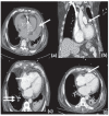
Case presentation: A 75-year-old Hispanic male presenting with dyspnea and diagnosed with cardiac tamponade is the subject of the study. Subsequent pericardiocentesis revealed biliary pericardial fluid (bilirubin of 7.6 mg/dl). The patient underwent extensive workup to identify a potential fistula between the hepatobiliary system and the pericardial space, which was non-revealing. The mechanism of bile entry into the pericardial space remains to be unidentified.
Literature review: A total of six previously published BPT were identified: all were males, with a mean age of 53.3 years (range: 31-73). Mortality was reported in two out of the six cases. The underlying etiology for pericardial tamponade varied across the cases: incidental pericardio-biliary fistula, traumatic pericardial injury, and presence of associated malignancy. - Conclusion: Biliary pericardial tamponade is a rare form of tamponade that warrants a prompt workup (e.g., Hepatobiliary Iminodiacetic Acid - HIDA scan) for an iatrogenic vs. traumatic pericardio- biliary fistula. As a first case in the literature, our case exhibits a biliary tamponade in the absence of an identifiable fistula.
Keywords: BPT; Pericardial effusion; biliary cardiac effusion; cardiac tamponade; pericardiobiliary fistula; pericardiocentesis..
Copyright© Bentham Science Publishers; For any queries, please email at epub@benthamscience.net.
Figures


References
-
- Marlatt S.W., Caride V.J., Prokop E.K. Incidental demonstration of pericardial fistula during hepatobiliary scintigraphy. J. Nucl. Med. 1991;32(3):518–520. - PubMed
-
- Chavez C., Golchian D. Donald Conn JF. Pericardiobiliary fistula: A rare complication of penetrating abdominal trauma. Appl. Radiol. 2018;47:24–26.
Publication types
MeSH terms
LinkOut - more resources
Full Text Sources
Other Literature Sources
Medical
Research Materials

