. 2020 Aug 3;217(8):e20192291.
doi: 10.1084/jem.20192291.

Tissue-resident memory T cell reactivation by diverse antigen-presenting cells imparts distinct functional responses

Affiliations

Tissue-resident memory T cell reactivation by diverse antigen-presenting cells imparts distinct functional responses

Jun Siong Low et al. J Exp Med. .

Abstract

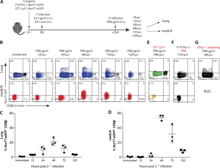
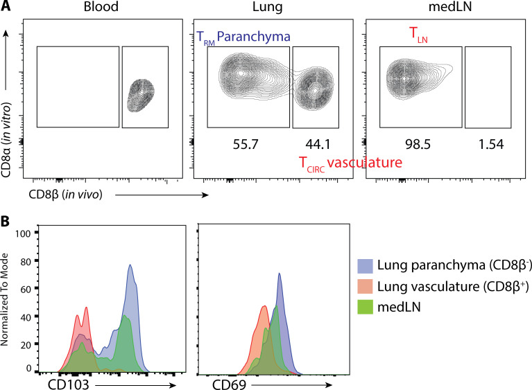
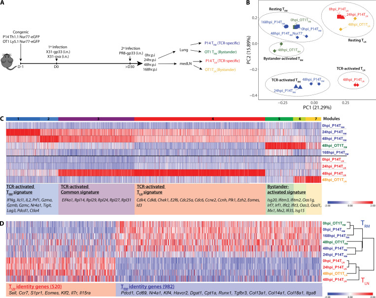
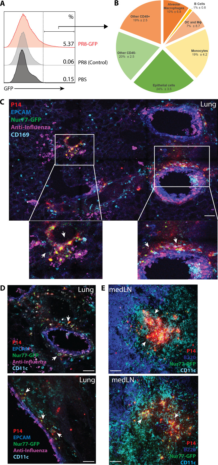
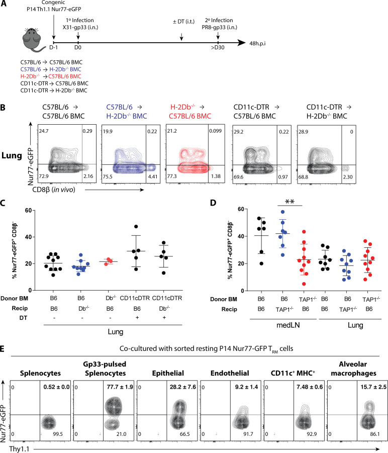
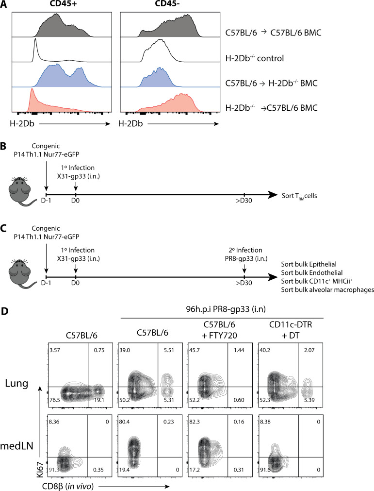
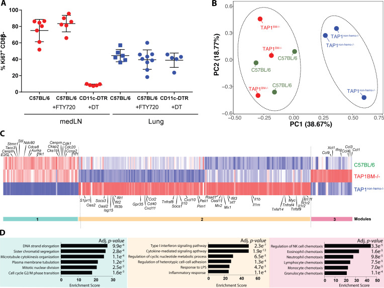
CD8+ tissue-resident memory T cells (TRM cells) are poised at the portals of infection and provide long-term protective immunity. Despite their critical roles, the precise mechanics governing TRM cell reactivation in situ are unknown. Using a TCR-transgenic Nur77-GFP reporter to distinguish "antigen-specific" from "bystander" reactivation, we demonstrate that lung CD8+ TRM cells are reactivated more quickly, yet less efficiently, than their counterparts in the draining LNs (TLN cells). Global profiling of reactivated memory T cells revealed tissue-defined and temporally regulated recall response programs. Unlike the reactivation of CD8+ TLN cells, which is strictly dependent on CD11c+XCR1+ APCs, numerous antigen-presenting partners, both hematopoietic and non-hematopoietic, were sufficient to reactivate lung CD8+ TRM cells, but the quality of TRM cell functional responses depended on the identity of the APCs. Together, this work uncovers fundamental differences in the activation kinetics, mechanics, and effector responses between CD8+ memory T cells in peripheral vs. lymphoid organs, revealing a novel tissue-specific paradigm for the reactivation of memory CD8+ T cells.


Conflict of interest statement

Disclosures: S.M. Kaech reported personal fees from Celsius Therapeutics and personal fees from the JEM Editorial Board outside the submitted work. No other disclosures were reported.

Figures

Figure 1.
Figure 1.
Lung TRM cells are reactivated more rapidly but less efficiently than TLN cells in medLNs. (A) P14/OT-I Nur77-GFP immune chimeras were generated by cotransferring naive P14+ (Thy1.1+) and OT-I+ (Ly5.1+) Nur77-GFP CD8+ T cells (5 × 10⁴ cells each) into C57BL/6 (Th1.2+/Ly5.2+) mice 1 d before i.n. coinfection with recombinant influenza X31-gp33 and X31-ova. 30 d later, the P14/OT-I Nur77-GFP immune chimeras were reinfected with PR8-gp33 i.n., and reactivation of P14+ lung TRM and medLN TLN cells was assessed at 0, 12, 24, 48, and 120 h.p.i. (B) Representative flow plots of Nur77-GFP expression in P14+ lung TRM cells and medLN TLN cells. In vivo labeling with αCD8β antibody was used to distinguish CD8+ TRM cells in the lung parenchyma from CD8+ T cells in the vasculature. (C and D) Frequency of Nur77-GFP+ CD8β P14+ cells (i.e., top left quadrant of flow plots) in B was quantified. Data are expressed as mean ± SD. (E and F) As negative controls, Nur77-GFP expression in OT-I+ and P14+ cells was examined after PR8-gp33 and PR8 2° infection, respectively. (G) To limit the contribution of TCIRC cells, a low dose of αThy1.1 (clone 19E12)–depleting antibody was administered i.p., and the frequency of Nur77-GFP+ P14+ TRM cell reactivation was examined. N.D., not detected. Antigen-activated P14+ lung TRM (blue) and medLN TLN (red) cells are distinguished from the bystander-activated OT-I+ lung TRM (green) and medLN TLN (gold) cell samples by color. Data shown are representative of two independent experiments (n = 3–5 mice/group).
Figure S1.
Figure S1.
Intravascular staining to distinguish parenchymal TRM cells from vasculature TLN cells. (A) Flu-immunized mice were injected i.v. with fluorescently labeled αCD8β antibody 5 min before sacrifice. Blood, lung, and medLN were collected and stained with αCD8α antibody ex vivo. Shown are gated on total CD8α+ T cells. (B) Expression of CD69 and CD103 in medLN, lung parenchyma (CD8a+ CD8β), and lung vasculature (CD8a+ CD8β+) are shown.
Figure S2.
Figure S2.
GzmB and CD98 are markers of bystander inflammation, and recruitment of circulating T cells is minimal at 48 h.p.i. (A and B) P14+ immunized mice were infected with PR8-gp33 (TCR-activation) or PR8 (bystander-activation) i.n., and the expression of GzmB (A) and CD98 (B) at 72 h.p.i. in the lung (top panel) and medLN (bottom panel) is shown. (C) P14+ TRM and TLN cells were sorted from resting P14+ chimera and co-cultured with control- or gp33-pulsed splenocytes, and Nur77-GFP expression was accessed 24 h later. (D and E) To limit the contribution of TCIRC cells to the readout of Nur77-GFP in cells in the lung, FTY720 (D) and a low dose of αThy1.1-depleting antibody (E) were administered i.p., and the effects on P14+ TRM cell reactivation was examined 48 h.p.i. n.s., not significant. Data are expressed as mean ± SD.
Figure 2.
Figure 2.
CD8+ memory T cells in the lung and medLNs display tissue-defined functional programs that are temporally regulated following reactivation in situ. (A) P14+ and OT-I+ memory CD8+ T cells were purified at 0, 24, 48, and 168 h after 2° infection from the lung and medLNs, and RNA-seq was performed. Activated P14+ T cells were sorted based on Nur77-GFP+ expression, and bystander-activated OT-I T cells were sorted based on CD98hi expression. (B) PCA of global gene expression showed five distinct clusters reflected by dotted ellipses. (C) Heatmap of differentially expressed TCR-activated genes and bystander-activated genes (log2FC ≥2 and P < 0.01). Seven modules were distinguished and several representative genes are shown. TCR-activated P14+ Nur77-GFP+ lung TRM (blue) and medLN TLN (red) are distinguished from the bystander-activated (Nur77-GFP CD98hi) OT-I+ lung TRM (green) and medLN TLN (gold) samples by color. Squares, 0 h.p.i; triangles, 24 h.p.i.; diamonds, 48 h.p.i; circles, 168 h.p.i. (D) TRM and TLN identity genes were identified by comparing 0 h resting P14+ memory CD8+ T cells from the lung and medLNs, based on 1.5-log2FC and P < 0.05, and the temporal changes of these identity genes following reactivation in TRM and TLN cell populations are shown in this heatmap.
Figure S3.
Figure S3.
TRM and TLN identity genes are maintained following reactivation. TRM and TLN identity genes were identified by comparing 0-h resting P14+ TRM cells with 0-h resting P14+ TLN cells based on 1.5-log2FC and P < 0.05, as represented in this volcano plot. 982 TRM identity genes and 520 TLN identity genes were identified.
Figure 3.
Figure 3.
Reactivated TRM cells are in close proximity to different infected cell types. (A) Influenza-immunized mice were infected with PR8-GFP and PR8 (as control), and the lungs were analyzed by flow cytometry 48 h.p.i. (B) The pie chart shows the types of infected GFP+ cells identified as a fraction of total GFP+ cells (pie chart: alveolar macrophages [CD45+ CD169+ SiglecF+], B cells [CD45+ B220+], DCs and macrophages [CD45+ SiglecF B220 CD11c+ Ly6C], monocytes [CD45+ SiglecF B220 CD11c+ Ly6C+], and epithelial cells [CD45 EPCAM+]). (C–E) P14+ immune chimeras were rechallenged with PR8-gp33, and the localization of activated P14+ Nur77-GFP+ cells in the lung (C and D) and medLN (E) at 48 h.p.i. was analyzed by immunofluorescence confocal imaging. Arrows indicate TCR-activated regions of P14+ Nur77-GFP+ cells and their interactions with infected cells and different immune subsets. Data shown are representative of two to three independent experiments (n = 3–5 mice/group). All scale bars indicate 50 µm.
Figure S4.
Figure S4.
Depletion validation and Zbtb46+ cells are required for medLN TLN cell, but not lung TRM cell, reactivation. P14+ immune chimeras were generated in various genetic hosts, and DT was administered i.t. 1 d before 2° infection to deplete DTR-expressing cells; for αGR-1 depletion, 200 μg αGR-1 (RB6-9C5) was administered i.p. daily starting the day before 2° rechallenge. (A) Validation of depletion strategies across different immunized-genetic hosts 48 h after the initial treatments in the lung or bronchoalveolar lavage. (B) Frequency of Nur77-GFP+ P14+ TRM cells in the lung and TLN cells in the medLN were quantified at 48 h.p.i. in Zbtb46-DTR hosts. Statistical analysis was performed using Student’s t test (two tailed); ****, P < 0.0001. (C) Flu viral titer was obtained by quantitative PCR of whole lung tissue for PR8 polymerase acidic protein (PA) 48 h.p.i. Data are expressed as mean ± SD.
Figure 4.
Figure 4.
CD11c+ XCR1+ cells are strictly required for medLN CD8+ TLN reactivation, but conventional APCs are dispensable for lung CD8+ TRM reactivation. (A) P14+ immune chimeras were generated in various genetic hosts as shown. For DTR mice, DT was administered i.t. 1 d before 2° infection to deplete DTR-expressing cells; for αGR-1 depletion, 200 μg αGR-1 (RB6-9C5) was administered i.p. daily starting the day before 2° rechallenge. For LCMV rechallenge experiments, flu-immunized mice were 2° infected with Armstrong strain of LCMV i.n. or LCMV-GFP. (B–D) Quantification of the frequency of Nur77-GFP+ P14+ TLN cells in the medLN (B) and P14+ TRM cells in the lung (C) at 48 h after 2° PR8-gp33 infection (B and C) or 48 h after 2° LCMV infection (D). Data shown are a collection of two or more independent experiments (n = 3–5 mice/group). (E and F) P14+ immune chimeras were rechallenged i.n. with LCMV-ZsGreen (E), and the colocalizations of ZsGreen signal with various immune cells in the medLN was quantified by Imaris (F). Top scale bars indicate 150 µm, and bottom scale bars indicate 20 µm. Data are expressed as mean ± SEM. Statistical analysis was performed using Student’s t test (two tailed) comparing immunized C57BL/6 to various treatment groups. ****, P < 0.0001.
Figure 5.
Figure 5.
CD8+ lung TRM cells can be reactivated by both hematopoietic and nonhematopoietic APCs. (A) P14+ immune chimeras were generated in different H-2Db−/− BMCs as outlined. For CD11c-DTR donor bone marrow groups, DT was administered 500 ng/mouse (i.p.) 1 d before 2° infection. (B) Representative flow plots of Nur77-GFP expression in the P14+ lung TRM cells at 48 h.p.i. and quantified in C. (D) P14+ immune chimeras were generated in TAP1−/− BMCs similar to A and the frequency of reactivated medLN TLN and lung TRM cells are quantified. (E) P14+ TRM cells were sorted from steady-state memory lung at >30 d p.i. and co-cultured with epithelial cells (EPCAM+), endothelial cells (CD31+), CD11c+ MHCII+ cells, and alveolar macrophages (CD169+) sorted from influenza-immunized mice that were 2°-reinfected with PR8-gp33 24 h prior. Flow plots of Nur77-GFP expression in P14+ TRM cells following overnight co-culture. Data shown are expressed as mean ± SD, representative of two independent experiments (n = 3–5 mice/group). Statistical analysis was performed using Student’s t test (two tailed). **, P < 0.01.
Figure S5.
Figure S5.
CD8+ lung TRM cells are reactivated by both hematopoietic and nonhematopoietic APCs, and consequentially, their 2° expansion in situ is independent of cDCs. (A) Validation of H-2Db expression in CD45+ and CD45 compartments of BMCs. (B) Schematic of ex vivo co-culture. P14+ TRM cells were sorted from steady-state memory lung at >30 d p.i. and co-cultured with epithelial cells (EPCAM+), endothelial cells (CD31+), CD11c+ MHCII+ cells, and alveolar macrophages (CD169+) sorted from influenza-immunized mice that were 2°-reinfected with PR8-gp33 24 h prior. (C) P14+ immune chimeras were generated in C57BL/6 or CD11c-DTR hosts and treated with either FTY720 (i.p., daily 2 days prior) or 100 ng DT (i.t., every 2–3 d) the day before 2° reinfection with PR8-GP33 i.n. Shown are representative flow plots of the expression of Ki67 on P14+ cells 96 h.p.i. (D) P14+ immune chimeras were generated in C57BL/6 or CD11c-DTR hosts and treated with either FTY720 (i.p.; daily 2 d prior) or 100 ng DT (i.t.; every 2–3 d) the day prior to 2o reinfection with PR8-GP33 i.n. 96 h.p.i. the expression of Ki67 on P14+ cells were examined and quantified. After infection., the expression of Ki67 on P14+ cells were examined and quantified.
Figure 6.
Figure 6.
Activation by hematopoietic and nonhematopoietic cells induces differential functional output in TRM cells. (A) P14+ immune chimeras were generated in C57BL/6 or CD11c-DTR hosts and treated with either FTY720 (i.p., daily 2 d prior) or 100 ng DT (i.t., every 2–3 d) the day before 2° reinfection with PR8-GP33 i.n. 96 h.p.i. the expression of Ki67 on P14+ cells was examined and quantified. Data are expressed as mean ± SEM. (B) Activated P14+ TRM cells were sorted based on Nur77-GFP+ expression in C57BL/6 TAP1+/+, TAP1BM−/−, TAP1nonhemo−/− BMCs, and RNA-seq was performed. PCA of global gene expression showed two distinct clusters reflected by dotted ellipses. (C) Heatmap of differentially expressed genes between the control and TAP1−/− BMCs (≥ or ≤1.5 log2FC and P < 0.05), filtered on all the differentially expressed genes of TCR and bystander TRM cell signature from Fig. 2 C. Three main modules were observed. (D) Genes from each module were parsed for enrichment of biological pathways using Enrichr. Top pathways from Gene Ontology Biological Processes with corresponding adjusted P values are shown. Relevant gene names are highlighted in heatmap in C.

References

    1. Ariotti S., Hogenbirk M.A., Dijkgraaf F.E., Visser L.L., Hoekstra M.E., Song J.-Y., Jacobs H., Haanen J.B., and Schumacher T.N.. 2014. T cell memory. Skin-resident memory CD8+ T cells trigger a state of tissue-wide pathogen alert. Science. 346:101–105. 10.1126/science.1254803 - DOI - PubMed
    1. De Baets S., Verhelst J., Van den Hoecke S., Smet A., Schotsaert M., Job E.R., Roose K., Schepens B., Fiers W., and Saelens X.. 2015. A GFP expressing influenza A virus to report in vivo tropism and protection by a matrix protein 2 ectodomain-specific monoclonal antibody. PLoS One. 10 e0121491 10.1371/journal.pone.0121491 - DOI - PMC - PubMed
    1. Ballesteros-Tato A., León B., Lund F.E., and Randall T.D.. 2010. Temporal changes in dendritic cell subsets, cross-priming and costimulation via CD70 control CD8(+) T cell responses to influenza. Nat. Immunol. 11:216–224. 10.1038/ni.1838 - DOI - PMC - PubMed
    1. Beura L.K., Mitchell J.S., Thompson E.A., Schenkel J.M., Mohammed J., Wijeyesinghe S., Fonseca R., Burbach B.J., Hickman H.D., Vezys V., et al. . 2018a Intravital mucosal imaging of CD8+ resident memory T cells shows tissue-autonomous recall responses that amplify secondary memory. Nat. Immunol. 19:173–182. 10.1038/s41590-017-0029-3 - DOI - PMC - PubMed
    1. Beura L.K., Wijeyesinghe S., Thompson E.A., Macchietto M.G., Rosato P.C., Pierson M.J., Schenkel J.M., Mitchell J.S., Vezys V., Fife B.T., et al. . 2018b T Cells in Nonlymphoid Tissues Give Rise to Lymph-Node-Resident Memory T Cells. Immunity. 48:327–338.e5. 10.1016/j.immuni.2018.01.015 - DOI - PMC - PubMed

Publication types