. 2020 Sep 9;28(3):411-421.e6.
doi: 10.1016/j.chom.2020.05.015. Epub 2020 Jun 10.

Clostridioides difficile Senses and Hijacks Host Heme for Incorporation into an Oxidative Stress Defense System

Affiliations

Clostridioides difficile Senses and Hijacks Host Heme for Incorporation into an Oxidative Stress Defense System

Reece J Knippel et al. Cell Host Microbe. .

Abstract

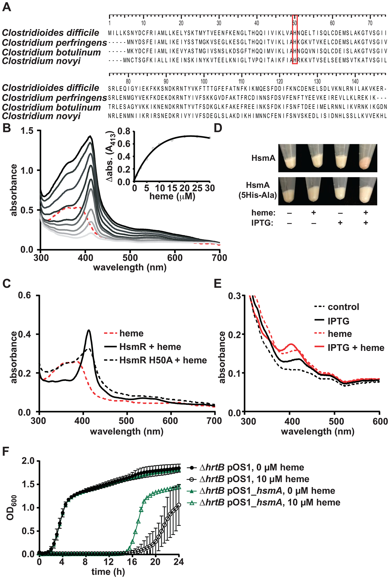
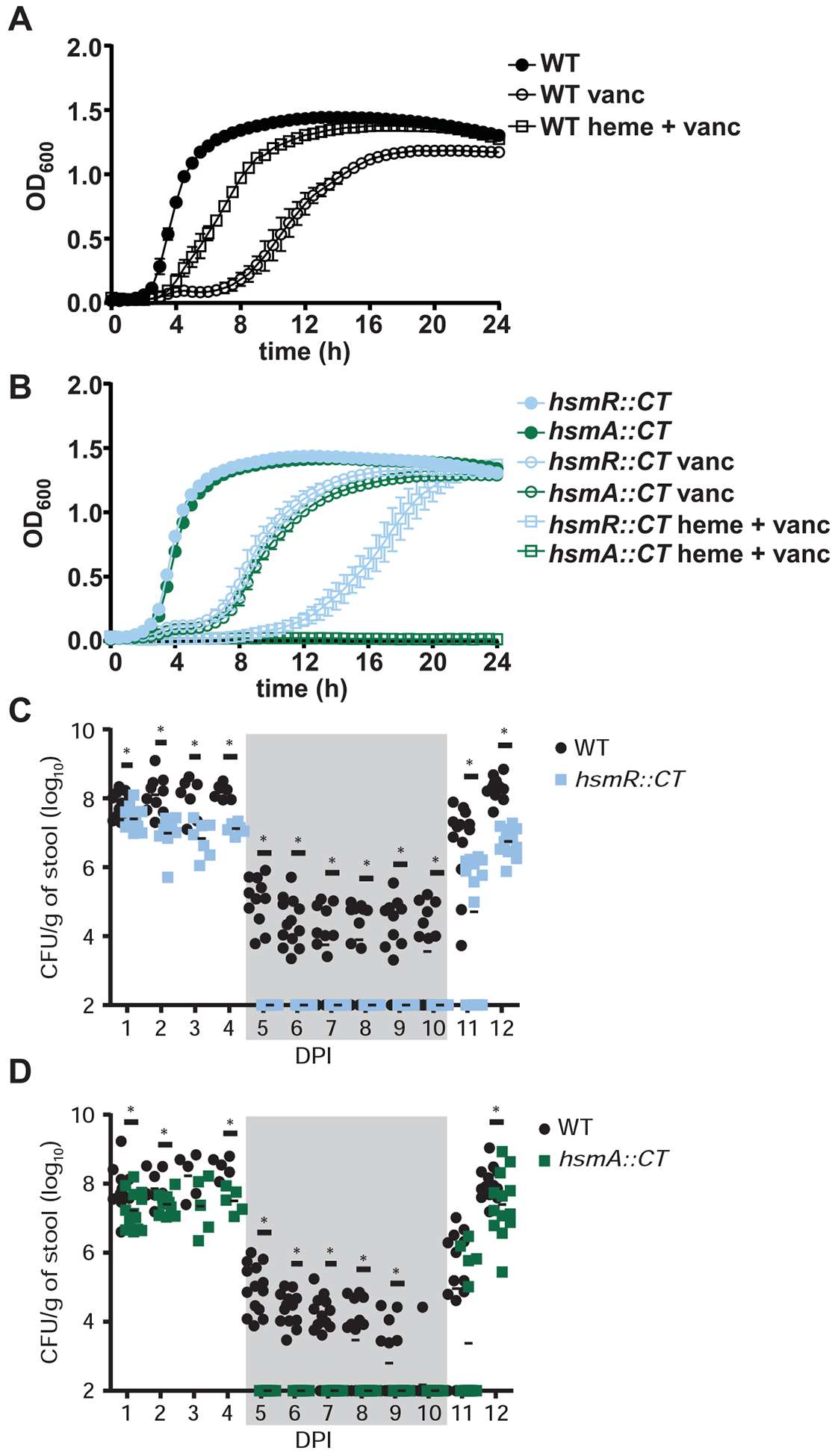
Clostridioides difficile infection of the colon leads to severe inflammation and damage to the gastrointestinal epithelium due to the production of potent toxins. This inflammatory tissue damage causes the liberation of high concentrations of host heme at infection sites. Here, we identify the C. difficile heme-sensing membrane protein system (HsmRA) and show that this operon induces a protective response that repurposes heme to counteract antimicrobial oxidative stress responses. HsmR senses vertebrate heme, leading to increased expression of the hsmRA operon and subsequent deployment of HsmA to capture heme and reduce redox damage caused by inflammatory mediators of protection and antibiotic therapy. Strains with inactivated hsmR or hsmA have increased sensitivity to redox-active compounds and reduced colonization persistence in a murine model of relapse C. difficile infection. These results define a mechanism exploited by C. difficile to repurpose toxic heme within the inflamed gut as a shield against antimicrobial compounds.

Keywords: Clostridioides difficile; antibiotic sensitivity; heme utilization; host-pathogen interactions; nutritional immunity; relapse infection.


Conflict of interest statement

Declaration of Interests Authors declare no competing interests.

Figures

Figure 1.
Figure 1.. hsmRA transcriptionally responds to and detoxifies heme.
(A) Schematic of the hsmRA operon. (B) hsmR and hsmA transcription determined by qRT-PCR. cDNA was reverse transcribed from RNA harvested from C. difficile R20291 grown in the presence of sodium hydroxide (NaOH, 500 μM), protoporphyrin IX (PPIX, 50 μM), iron sulfate (50 μM) or heme (50 μM). (C) hsmR, hsmA, hatR, and hatT transcription determined by qRT-PCR of cDNA reverse transcribed from RNA harvested from C. difficile grown in the presence of a low concentration range of heme (0.25 – 1 μM). Transcription is graphed as the fold change relative to an untreated control. The data are a representative of three independent experiments with standard deviation shown. Statistical significance was determined using the multiple comparison two-way ANOVA test with the Sidak correction for multiple comparisons * denotes p < 0.05. (D) Growth of WT, hsmR::CT, and hsmA::CT strains in the presence or absence of heme (25 μM). The data are a representative from three independent experiments each in biological triplicate with standard error of the mean. See also Figure S1 and Tables S1 and S2.
Figure 2.
Figure 2.. HsmR binds and senses heme and HsmA reduces heme toxicity through sequestration.
(A) Alignment of HsmR homologues in other pathogenic clostridial species. Red box denotes conserved histidine residue. (B) Absorption spectra of heme binding to recombinant HsmR. Increasing concentrations of heme (1 to 30 μM) were added to 10 μM protein. The spectrum corresponding to 10 μM heme is shown as a dashed red line. HsmR with increasing concentrations of heme are shown as gray lines. The inset displays change in absorbance at 413 nm for HsmR bound to heme minus the corresponding heme alone peak. (C) Absorption spectra of 10 μM heme binding to HsmR or HsmR H50A. (D) E. coli pET15b_hsmA or pET15b_hsmA_5His-Ala cell pellets in the presence or absence of heme (10 μM) and IPTG (1 mM). (E) Absorption spectra of solubilized membrane fractions of the cell pellets from D. (F) Growth of S. aureus ΔhrtB pOS1 and pOS1_hsmA strains in the presence or absence of heme (10 μM). The data are a representative from three independent experiments with standard error of the mean. See also Figures S2, S3, and S4 and Tables S1 and S2.
Figure 3.
Figure 3.. HsmR is an activator of the hsmRA operon.
(A) hsmR and hsmA transcription determined by qRT-PCR. cDNA was reverse transcribed from RNA harvested WT or hsmR::CT grown to early exponential phase (0.3 abs) and exposed to heme (50 μM) for 30 min. Data are represented as fold change relative to untreated WT. (B) Electrophoretic mobility shift assay (EMSA) demonstrates direct and specific DNA binding by HsmR to the 5’ UTR of hsmR, but not the 5’ UTR of the unrelated gene CDR20291_0783 (see Figure 1A). (C) RNA-sequencing analysis comparing RNA from heme treated (25 μM for 30 min) WT to an untreated WT control. (D) RNA-sequencing analysis comparing RNA from heme treated (25 μM for 30 min) hsmR::CT to an untreated hsmR::CT control. Dashed lines represent genes of fold change > 2. Samples with p-value > 1 × 10⁻⁵ are represented as 5 on the graph. Solid black line denotes p < 0.05. Statistical significance was determined using the multiple comparison two-way ANOVA test with the Sidak correction for multiple comparisons comparing the means of each group to one another. * denotes p-value < 0.05, n.s. denotes not significant. See also Figure S1 and Tables S1, S3, S4 and S5.
Figure 4.
Figure 4.. HsmA reduces oxidative stress.
(A) C. difficile (CD) WT and hsmA::CT strains were grown for 6 hours followed by treatment with or without heme (25 μM) for 30 min. Samples were exposed to atmospheric oxygen and oxidative stress generation was determined by measuring fluorescence of dihydrorhodamine 123 (DHR123; ex. 507 nm, em. 529). (B) Growth of S. aureus (SA) ΔΔsod pOS1 and pOS1_hsmA strains in the presence or absence of paraquat (2 mM) and dihydrorhodamine 123. (C) Oxidative stress generation was quantified by measuring fluorescence of DHR123 from SA ΔΔsod pOS1 (empty vector), SA ΔΔsod pOS1_hsmA, or SA ΔΔsod pOS1_hsmA_5His-Ala (point mutant defective for heme binding) in the presence or absence of paraquat. The data are a representative from three independent experiments with standard error of the mean. (D) S. aureus ΔΔsod strains carrying the empty pOS1 vector or pOS1_hsmA were co-incubated with neutrophils derived from murine bone marrow (MOI of 1) for 12 hours. S. aureus CFUs were enumerated and compared to untreated controls. Statistical significance was determined using the multiple comparison two-way ANOVA test with the Sidak correction for multiple comparisons comparing the means of each group to one another. * denotes p-value < 0.05. See also Figure S4 and Table S2.
Figure 5.
Figure 5.. The hsmRA operon decreases sensitivity to vancomycin in the presence of heme during infection.
(A, B) Growth of WT, hsmR::CT and hsmA::CT in the presence or absence of vancomycin (100 μg/mL) and heme (10 μM). The data are a representative from three independent experiments with standard error of the mean. (C, D) CFU analysis of mice coinfected with WT and hsmR::CT or WT and hsmA::CT strains for 12 days post infection (DPI) with standard error of the mean (n=14/group). Vancomycin treatment (0.4 mg/mL) was administered on days 5–10 (denoted by gray shading) and removed on day 11. The data are presented with standard error of the mean. Statistical significance was determined using the multiple comparison two-way ANOVA test with the Bonferroni correction for multiple comparisons comparing the means of each group to one another. * denotes p-value < 0.05. See also Figure S4 and S5.
Figure 6.
Figure 6.. C. difficile utilization of host heme for protection against oxidative stress.
(A) Graphical representation of the C. difficile host pathogen interface during infection. Toxin mediated inflammation induces translocation and lysis of erythrocyte in the gastrointestinal lumen resulting in high concentrations of heme. (B) Host heme is sensed by HsmR and incorporated into HsmA, providing protection against oxidative stress produced by host immune cells and environment. Concurrently, HatR binds heme derepressing the hatRT operon and leading to subsequent efflux of heme by HatT.

Comment in

References

    1. Abt MC, McKenney PT, and Pamer EG (2016). Clostridium difficile colitis: pathogenesis and host defence. Nat Rev Microbiol. - PMC - PubMed
    1. Asard H, Barbaro R, Trost P, and Bérczi A (2013). Cytochromes b561: ascorbate-mediated trans-membrane electron transport. Antioxid Redox Signal 19, 1026–1035. - PMC - PubMed
    1. Attia AS, Benson MA, Stauff DL, Torres VJ, and Skaar EP (2010). Membrane damage elicits an immunomodulatory program in Staphylococcus aureus. PLoS Pathog 6, e1000802. - PMC - PubMed
    1. Benjamini Y, and Hochberg Y (1995). Controlling the False Discovery Rate: A Practical and Powerful Approach to Multiple Testing. Journal of the Royal Statistical Society Series B (Methodological) 57, 289–300.
    1. Blattner FR, Plunkett G, Bloch CA, Perna NT, Burland V, Riley M, Collado-Vides J, Glasner JD, Rode CK, Mayhew GF, et al. (1997). The complete genome sequence of Escherichia coli K-12. Science 277, 1453–1462. - PubMed

Publication types

MeSH terms

LinkOut - more resources