. 2020 Sep 2;28(9):1974-1986.
doi: 10.1016/j.ymthe.2020.05.021. Epub 2020 May 30.

Targeting CD33 in Chemoresistant AML Patient-Derived Xenografts by CAR-CIK Cells Modified with an Improved SB Transposon System

Affiliations

Targeting CD33 in Chemoresistant AML Patient-Derived Xenografts by CAR-CIK Cells Modified with an Improved SB Transposon System

Maria Caterina Rotiroti et al. Mol Ther. .

Abstract

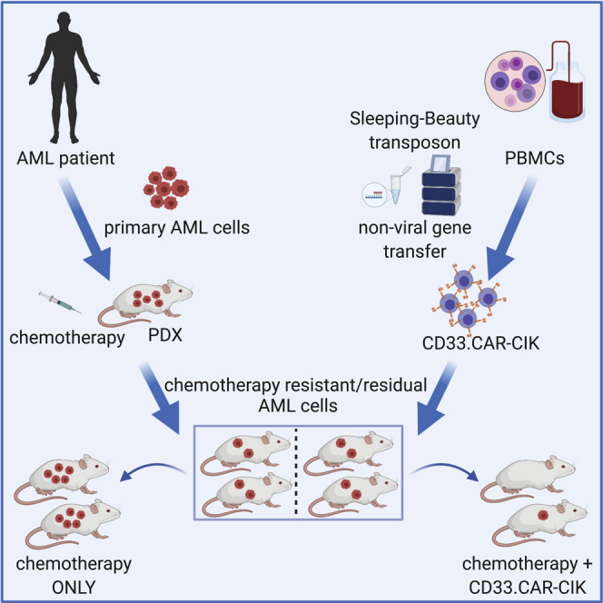
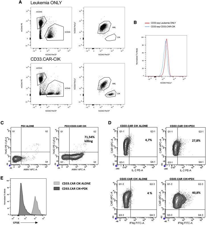
The successful implementation of chimeric antigen receptor (CAR)-T cell therapy in the clinical context of B cell malignancies has paved the way for further development in the more critical setting of acute myeloid leukemia (AML). Among the potentially targetable AML antigens, CD33 is insofar one of the main validated molecules. Here, we describe the feasibility of engineering cytokine-induced killer (CIK) cells with a CD33.CAR by using the latest optimized version of the non-viral Sleeping Beauty (SB) transposon system "SB100X-pT4." This offers the advantage of improving CAR expression on CIK cells, while reducing the amount of DNA transposase as compared to the previously employed "SB11-pT" version. SB-modified CD33.CAR-CIK cells exhibited significant antileukemic activity in vitro and in vivo in patient-derived AML xenograft models, reducing AML development when administered as an "early treatment" and delaying AML progression in mice with established disease. Notably, by exploiting an already optimized xenograft chemotherapy model that mimics human induction therapy in mice, we demonstrated for the first time that CD33.CAR-CIK cells are also effective toward chemotherapy resistant/residual AML cells, further supporting its future clinical development and implementation within the current standard regimens.

Keywords: AML; CAR; CD33; Sleeping Beauty transposon; cytokine-induced killer cells; immunotherapy; non-viral gene transfer.


Figures

None
Graphical abstract
Figure 1
Figure 1
CAR Expression, Integration Site Analysis, and SB Transposase Detection in SB-Modified CD33.CAR-CIK Cells at Day 21 (A) Percentage of CAR expression. (B) MFI values. (C) The integration distribution of SB represented on the chromosome level for each healthy donor, where healthy donor 1’s 8,063 integrations are in red, donor 2’s 7,213 are in blue, and donor 3’s 6,856 integrations are in green. (D) Pie chart of SB integrations in relation to RefSeq genome annotations as a percentage of the total 22,132 integrations. The integrations within 1 kb upstream and downstream of TSS, 3′, and 5′ UTR were also considered. 94.5% of the integrations were intergenic or in introns. (E) SB transposase transcripts. (F) Western blotting for SB100X in three different CD33.CAR-CIK cell productions. The box contains data that fall between the first and third quartiles, the horizontal line indicates the median, the diamond indicates the mean, and the brackets delineate 1.5 times the interquartile range. Individual data are also shown. Comparisons between groups were done according to Wilcoxon rank sum test.
Figure 2
Figure 2
In Vitro Characterization of SB-Modified CD33.CAR-CIK Cells (A) Short-term cytotoxic assay E:T 5:1 (THP-1, n = 7 for NO DNA and SB11/pT, n = 3 for SB100X/pT4; MA9, n = 7 for NO DNA and SB11/pT, n = 6 for SB100X/pT4; PRIMARY AML, n = 6; MHH-CALL-4, n = 7 for NO DNA and SB11/pT, n = 4 for SB100X/pT4). (B and C) Intracellular staining for IFN-γ (B) and IL-2 (C) after 5 h co-culture of CIK cells with the various targets E:T 1:3 (THP-1, n = 7 for NO DNA and SB11/pT, n = 3 for SB100X/pT4; MA9, n = 7 for NO DNA and SB11/pT, n = 4 for SB100X/pT4; PRIMARY AML, n = 7; MHH-CALL-4, n = 7 for NO DNA and SB11/pT, n = 3 for SB100X/pT4). (D) Intracellular staining for Ki67 after 72 h co-culture of CIK cells with the various targets, E:T 1:1 (THP-1, n = 7 for NO DNA and SB11/pT, n = 3 for SB100X/pT4; MA9, n = 3 for NO DNA and SB11/pT, n = 2 for SB100X/pT4; PRIMARY AML, n = 7 for NO DNA and SB11/pT, n = 2 for SB100X/pT4; MHH-CALL-4, n = 7 for NO DNA and SB11/pT, n = 3 for SB100X/pT4). (E and F) CFSE proliferation assay of SB11/pT- (E) and SB100X/pT4- (F) engineered CD33.CAR-CIK cells after 72 h co-culture with the various targets, E:T 1:1. The result of a representative experiment out of 3 is shown. Data represented are the result of mean ± SEM; comparisons between groups were done according to the Kruskal-Wallis test.
Figure 3
Figure 3
In Vivo Antitumor Efficacy of SB100X/pT4 CD33.CAR-CIK Cells against OCI-AML3 Luc-GFP Cell Line (A) NSG mice were inoculated with 106 OCI-AML3 Luc-GFP cells. Starting from day 3, mice received once weekly, for 3 weeks, 107 CD33.CAR-CIK cells. (B) Bioluminescence imaging was performed using the IVIS lumina III imaging system (Perkin-Elmer). Tumor burden visualized on days 3, 10, 17, and 24. (C) Kaplan-Meier survival curve representing survival outcomes. Statistical significance was calculated with a Mantel-Cox (log rank) test, p = 0.0082.
Figure 4
Figure 4
In Vivo Antitumor Efficacy of SB100X/pT4 CD33.CAR-CIK Cells in an “Early Treatment” Model (A) NSG mice were inoculated with 106 cells of a secondary PDX. Starting from day 5, mice received once weekly, for 3 weeks, 107 CD33.CAR-CIK cells. Mice were sacrificed 7 days after the last injection. (B–D) Percentage of leukemia engraftment in the peripheral blood (PB) (B), in the bone marrow (BM) (C), and spleen (D). (E) Percentage of CD3+ cells in the different districts detected in each of the treated mouse at the end of the experiment. (F) A representative plot of the BM from each group is shown. Comparisons between groups were done according to Wilcoxon rank sum test.
Figure 5
Figure 5
CD33 Expression on AML Cells and Ex Vivo Treatment of Residual Leukemic Cells (A and B) Representative dot plots (A) and histogram (B) showing CD33 antigen expression on AML-PDX in untreated (leukemia ONLY) and CD33.CAR-CIK cell-treated mice (BM). (C) Representative dot plots showing the staining with annexin V and 7AAD on ex vivo leukemic cells alone and in the presence of CD33.CAR-CIK cells. (D) Representative dot plots of cytokine production by CD33.CAR-CIK cells following target stimulation. (E) CD33.CAR-CIK cell proliferation by CFSE dilution following target stimulation.
Figure 6
Figure 6
In Vivo Antitumor Efficacy of SB100X/pT4 CD33.CAR-CIK Cells in an “AML Treatment” Model (A) NSG mice were inoculated with 106 cells of a secondary PDX. When a substantial tumor burden was evident in the BM, mice received once weekly, for 3 weeks, 107 CD33.CAR-CIK cells. Mice were sacrificed 7 days after the last injection. (B) Representative dot plot of BM engraftment before treatment. (C–E) Percentage of leukemia engraftment in the PB (C), the BM (D), and spleen (E). (F) Percentage of CD3+ cells in the different districts detected in each of the treated mouse at the end of the experiment. (G) A representative dot plot of CD3 and CAR expression from PB of a treated mouse is shown. Comparisons between groups were done according to Wilcoxon rank sum test.
Figures 7
Figures 7
In Vivo Antitumor Efficacy of SB100X/pT4 CD33.CAR-CIK Cells in the Xenograft Chemotherapy Model (A) NSG mice were inoculated with 106 cells of a secondary PDX. When a substantial tumor burden was evident in the BM (≈20%), mice received the “5+3” chemotherapy induction protocol resulting in a remission-like status with disease recurrence within short time. At the first evidence of disease recurrence, mice received once weekly, for 3 weeks, 107 CD33.CAR-CIK cells. Mice were sacrificed 7 days after the last injection. (B) Percentage of leukemia engraftment in the BM of chemotherapy-only-treated mice (CONTROL) and CD33.CAR-CIK cells treated mice before chemotherapy (CTX), after CTX and after CD33.CAR-CIK cell treatment. (C and D) Percentage of leukemia engraftment at sacrifice in PB (C) and spleen (D). (E) Percentage of CD3+ cells in the different districts detected in each of the treated mouse at the end of the experiment. Comparisons between groups were done according to Wilcoxon rank sum test.

References

    1. Gross G., Waks T., Eshhar Z. Expression of immunoglobulin-T-cell receptor chimeric molecules as functional receptors with antibody-type specificity. Proc. Natl. Acad. Sci. USA. 1989;86:10024–10028. - PMC - PubMed
    1. Maude S.L., Laetsch T.W., Buechner J., Rives S., Boyer M., Bittencourt H., Bader P., Verneris M.R., Stefanski H.E., Myers G.D. Tisagenlecleucel in children and young adults with B-cell lymphoblastic leukemia. N. Engl. J. Med. 2018;378:439–448. - PMC - PubMed
    1. Neelapu S.S., Locke F.L., Bartlett N.L., Lekakis L.J., Miklos D.B., Jacobson C.A., Braunschweig I., Oluwole O.O., Siddiqi T., Lin Y. Axicabtagene ciloleucel CAR T-cell therapy in refractory large B-Cell lymphoma. N. Engl. J. Med. 2017;377:2531–2544. - PMC - PubMed
    1. Walter R.B., Appelbaum F.R., Estey E.H., Bernstein I.D. Acute myeloid leukemia stem cells and CD33-targeted immunotherapy. Blood. 2012;119:6198–6208. - PMC - PubMed
    1. Ehninger A., Kramer M., Röllig C., Thiede C., Bornhäuser M., von Bonin M., Wermke M., Feldmann A., Bachmann M., Ehninger G., Oelschlägel U. Distribution and levels of cell surface expression of CD33 and CD123 in acute myeloid leukemia. Blood Cancer J. 2014;4:e218. - PMC - PubMed

Publication types

MeSH terms