. 2020 Jun 9;21(11):4112.
doi: 10.3390/ijms21114112.

Ablation of the miRNA Cluster 24 Has Profound Effects on Extracellular Matrix Protein Abundance in Cartilage

Affiliations

Ablation of the miRNA Cluster 24 Has Profound Effects on Extracellular Matrix Protein Abundance in Cartilage

Veronika S Georgieva et al. Int J Mol Sci. .

Abstract

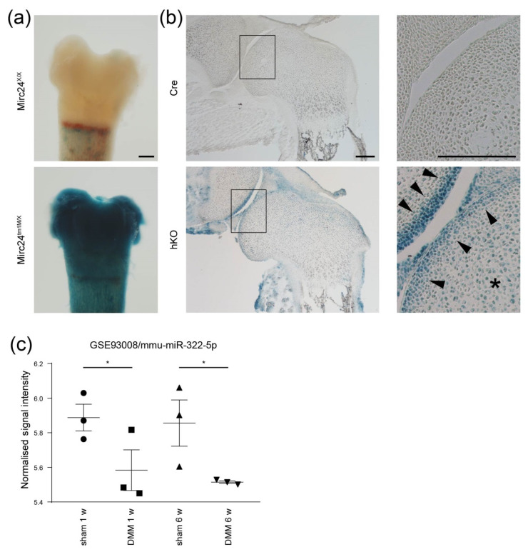
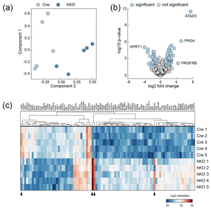
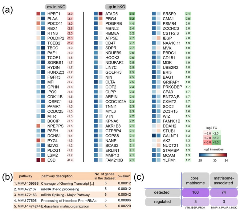
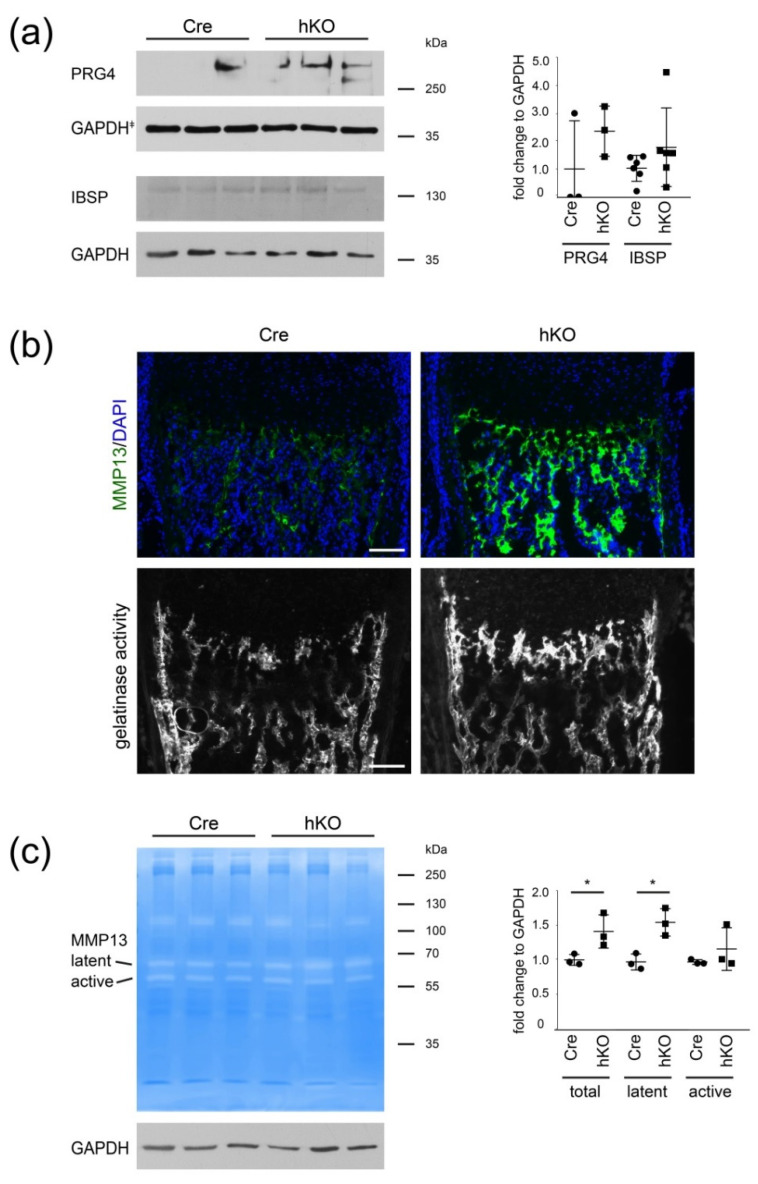
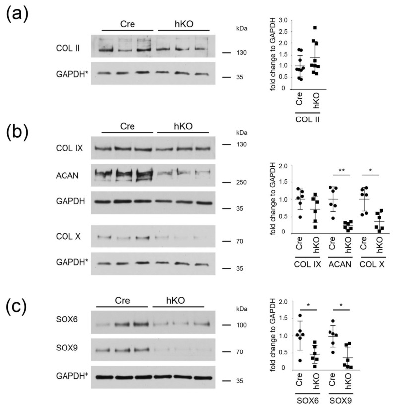
MicroRNAs (miRNAs) regulate cartilage differentiation and contribute to the onset and progression of joint degeneration. These small RNA molecules may affect extracellular matrix organization (ECM) in cartilage, but for only a few miRNAs has this role been defined in vivo. Previously, we showed that cartilage-specific genetic ablation of the Mirc24 cluster in mice leads to impaired cartilage development due to increased RAF/MEK/ERK pathway activation. Here, we studied the expression of the cluster in cartilage by LacZ reporter gene assays and determined its role for extracellular matrix homeostasis by proteome and immunoblot analysis. The cluster is expressed in prehypertrophic/hypertrophic chondrocytes of the growth plate and we now show that the cluster is also highly expressed in articular cartilage. Cartilage-specific loss of the cluster leads to increased proteoglycan 4 and matrix metallopeptidase 13 levels and decreased aggrecan and collagen X levels in epiphyseal cartilage. Interestingly, these changes are linked to a decrease in SRY-related HMG box-containing (SOX) transcription factors 6 and 9, which regulate ECM production in chondrocytes. Our data suggests that the Mirc24 cluster is important for ECM homoeostasis and the expression of transcriptional regulators of matrix production in cartilage.

Keywords: MMP13; Mirc24; PRG4; SOX6; SOX9; articular; cartilage; extracellular matrix; miR-322; miR-503.


Conflict of interest statement

The authors declare no conflict of interest.

Figures

Figure 1
Figure 1
Characterization of the Mirc24 cluster expression in epiphyseal cartilage. Expression of the LacZ reporter gene in (a) isolated femora of Mirc24X/X and Mirc24tm1M/X female newborns and (b) sections of the distal epiphyseal femoral joints of Col2a1-Cre (Cre) and Col2a1-Cre-Mirc24 tm1M/Y (hKO) newborn males was detected by X-Gal staining and assessed by microscopy. Asterix: epihyseal head cartilage, arrowheads: articular cartilage, bar: (a,b) 200 µm. (c) Expression of the Mirc24 cluster-encoded miR-322-5p in articular cartilage from mice subjected to sham or destabilization of the medial meniscus (DMM) surgery (n = 3) at 1 and 6 weeks was determined by Geo2R analysis and expression intensity and p-value of the GEO dataset GSE93008 are shown.
Figure 2
Figure 2
Proteome analysis of epiphyseal cartilage from Mirc24 deficient mice. (a) Principal component analysis of the proteome dataset. Each circle represents an individual sample of epiphyseal cartilage isolated from Col2a1-Cre (Cre) or Col2a1-Cre-Mirc24tm1M/Y (hKO) newborn mice. (b) Volcano plot illustrating significant changes within the dataset. Proteins with the highest fold change are marked (blue). (c) Non-averaged hierarchical clustered intensity plot (distant metrics—euclidean, linkage rule—complete) of differentially abundant proteins. Protein symbols within the clusters are given and the relative protein abundance is indicated (blue—low, red—high). Proteins with the highest fold change are marked (blue) (arrowheads).
Figure 3
Figure 3
Identification of regulated protein interaction networks in cartilage extracts from Mirc24 deficient mice. (a) Proteins with decreased (dw in hKO, left) and increased (up in hKO, right) intensity are ranked according to the fold change. The results of the (b) reactome pathway characterization of increased proteins within string database and the (c) matrisome analysis of regulated entities are shown.
Figure 4
Figure 4
Characterization of PRG4, IBSP and MMP13 levels in femoral joint cartilage. (a) PRG4 and IBSP protein levels were determined in newborn cartilage extracts from Col2a1-Cre (Cre) and Col2a1-Cre-Mirc24tm1M/Y (hKO) mice using immunoblots. GAPDH was used as loading control. Quantification is shown (graph, PRG4 n = 3, IBSP n = 6). GAPDH indicates that PRG4, SOX6 and SOX9 were tested on a single blot and the same GAPDH blot is shown in Figure 4a and Figure 5c. Additional immunoblots are shown in Supplemental Figure S3a. (b) MMP13 localization was studied by immunofluorescence analysis (DAPI—blue, MMP13—green, pseudocolored) and gelatinase activity was determined by in situ zymography in newborn femoral epiphysis. Brightness was adjusted for visualization. (c) MMP13 activity in femoral extracts was analyzed by gelatin zymography. Latent proMMP13 and active MMP13 are indicated. GAPDH was used as loading control. Fold change of total, latent and active MMP13 in hKO extracts compared to Cre was determined (graph, n = 3). Molecular weights of Thermo Scientific™ PageRuler™ Plus Prestained 10–250 kDa Protein Ladder bands are given. Scale bar 100 µm. p < 0.05 (*).
Figure 5
Figure 5
Analysis of the ECM and SOX protein expression in cartilage. Cartilage extracts from Col2a1-Cre (Cre) and Col2a1-Cre-Mirc24tm1M/Y (hKO) mice were analyzed for the presence of (a) collagen II, (b) collagen IX, aggrecan, and X as well as (c) SOX6 and SOX9 using immunoblots. GAPDH was used as loading control. GAPDH* indicates that Collagen II and X were tested on a single blot and the same GAPDH blot is shown in (a) and (b). GAPDH indicates that PRG4, SOX6 and SOX9 were tested on a single blot and the same GAPDH blot is shown in Figure 4a and Figure 5c. Quantification is shown (graph, n ≥ 6). Additional immunoblots are shown in Supplemental Figure S3b–d. Molecular weights of Thermo Scientific™ PageRuler™ Plus Prestained 10–250 kDa Protein Ladder bands are given. p < 0.05 (*), p < 0.01 (**).

References

    1. Endisha H., Rockel J., Jurisica I., Kapoor M. The complex landscape of microRNAs in articular cartilage: Biology, pathology, and therapeutic targets. JCI insight. 2018;3:e121630. doi: 10.1172/jci.insight.121630. - DOI - PMC - PubMed
    1. Kung L.H., Zaki S., Ravi V., Rowley L., Smith M.M., Bell K.M., Bateman J.F., Little C.B. Utility of circulating serum miRNAs as biomarkers of early cartilage degeneration in animal models of post-traumatic osteoarthritis and inflammatory arthritis. Osteoarthr. Cartil. 2017;25:426–434. doi: 10.1016/j.joca.2016.09.002. - DOI - PubMed
    1. Miyaki S., Sato T., Inoue A., Otsuki S., Ito Y., Yokoyama S., Kato Y., Takemoto F., Nakasa T., Yamashita S., et al. MicroRNA-140 plays dual roles in both cartilage development and homeostasis. Genes Dev. 2010;24:1173–1185. doi: 10.1101/gad.1915510. - DOI - PMC - PubMed
    1. Aszodi A., Bateman J.F., Gustafsson E., Boot-Handford R., Fassler R. Mammalian skeletogenesis and extracellular matrix: What can we learn from knockout mice? Cell Struct. Funct. 2000;25:73–84. doi: 10.1247/csf.25.73. - DOI - PubMed
    1. Etich J., Holzer T., Pitzler L., Bluhm B., Brachvogel B. MiR-26a modulates extracellular matrix homeostasis in cartilage. Matrix. Biol. 2015;43:27–34. doi: 10.1016/j.matbio.2015.02.014. - DOI - PubMed

MeSH terms