

Update on vasculitis: overview and relevant dermatological aspects for the clinical and histopathological diagnosis - Part II
- PMID: 32527591
- PMCID: PMC7335877
- DOI: 10.1016/j.abd.2020.04.004
Update on vasculitis: overview and relevant dermatological aspects for the clinical and histopathological diagnosis - Part II
Abstract
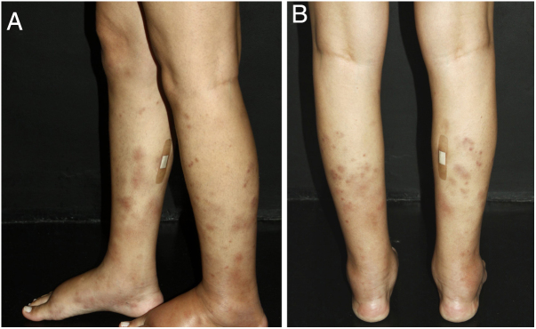
Vasculitis is a group of several clinical conditions in which the main histopathological finding is fibrinoid necrosis in the walls of blood vessels. This article assesses the main dermatological aspects relevant to the clinical and laboratory diagnosis of small- and medium-vessel cutaneous and systemic vasculitis syndromes. The most important aspects of treatment are also discussed.
Keywords: Anti-neutrophil cytoplasmic antibodies; Churg-Strauss syndrome; Henoch-Schönlein purple; Leukocytoclastic cutaneous vasculitis; Systemic vasculitis; Vasculitis; Vasculitis associated with lupus of the central nervous system.
Copyright © 2020 Sociedade Brasileira de Dermatologia. Published by Elsevier España, S.L.U. All rights reserved.
Figures
References
-
- Carlson J.A. The histological assessment of cutaneous vasculitis. Histopathology. 2010;56:3–23. - PubMed
-
- Demirkesen C. Approach to cutaneous vasculitides with special emphasis on small vessel vasculitis. Curr Opin Rheumatol. 2017;29:39–44. - PubMed
-
- Chen K.R., Carlson J.A. Clinical approach to cutaneous vasculitis. Am J Clin Dermatol. 2008;9:71–92. - PubMed
-
- Micheletti R.G. Small vessel vasculitis of the skin. In: Dammacco F., Ribatti D., Vacca A., editors. Systemic vasculitides: current status and perspectives. Springer International Publishing; Cham: 2016. pp. 233–244.
-
- Zanoni G., Girolomoni G., Bonetto C., Trotta F., Häusermann P., Opri R. Single organ cutaneous vasculitis: case definition & guidelines for data collection, analysis, and presentation of immunization safety data. Vaccine. 2016;34:6561–6571. - PubMed
Publication types
MeSH terms
LinkOut - more resources
Full Text Sources
Medical
