. 2020 Jun 11;12(12):11754-11767.
doi: 10.18632/aging.103341. Epub 2020 Jun 11.

miR-107 inhibition upregulates CAB39 and activates AMPK-Nrf2 signaling to protect osteoblasts from dexamethasone-induced oxidative injury and cytotoxicity

Affiliations

miR-107 inhibition upregulates CAB39 and activates AMPK-Nrf2 signaling to protect osteoblasts from dexamethasone-induced oxidative injury and cytotoxicity

Yu Zhuang et al. Aging (Albany NY). .

Abstract

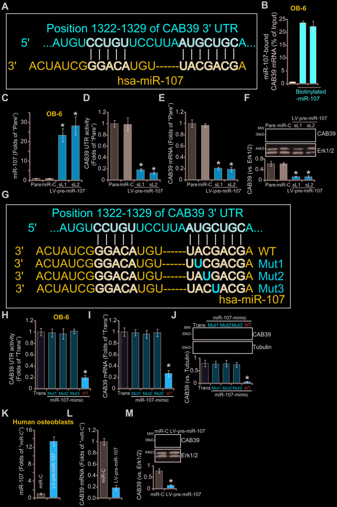
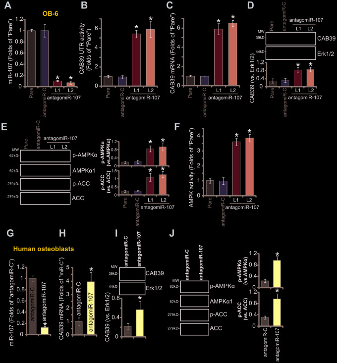
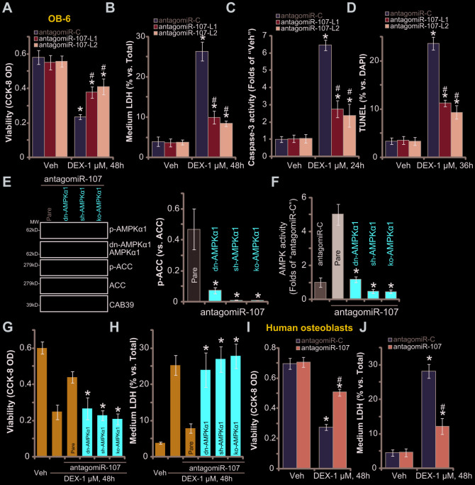
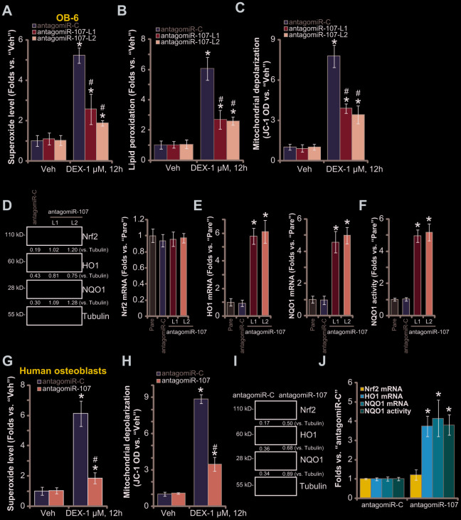
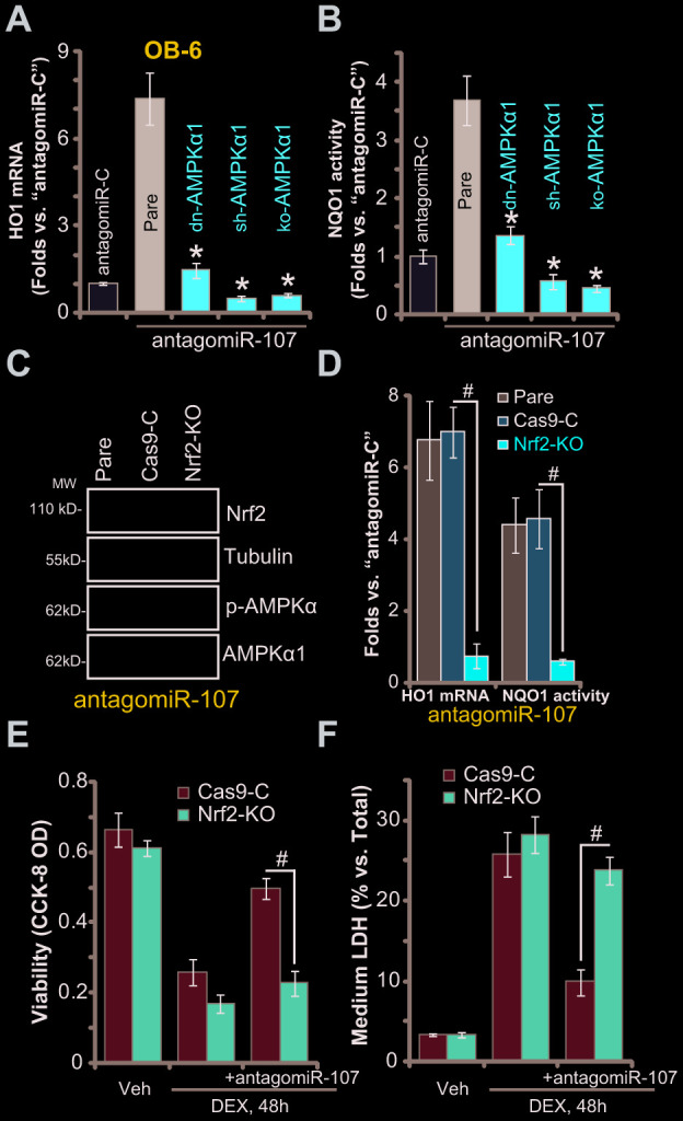
To human osteoblasts dexamethasone (DEX) treatment induces significant oxidative injury and cytotoxicity. Inhibition of CAB39 (calcium binding protein 39)-targeting microRNA can induce CAB39 upregulation, activating AMP-activated protein kinase (AMPK) signaling and offering osteoblast cytoprotection. Here we identified a novel CAB39-targeting miRNA: the microRNA-107 (miR-107). RNA-Pull down assay results demonstrated that the biotinylated-miR-107 directly binds to CAB39 mRNA in OB-6 human osteoblastic cells. Forced overexpression of miR-107, by infection of pre-miR-107 lentivirus or transfection of wild-type miR-107 mimic, largely inhibited CAB39 expression in OB-6 cells and primary human osteoblasts. Contrarily, miR-107 inhibition, by antagomiR-107, increased its expression, resulting in AMPK cascade activation. AntagomiR-107 largely attenuated DEX-induced cell death and apoptosis in OB-6 cells and human osteoblasts. Importantly, osteoblast cytoprotection by antagomiR-107 was abolished with AMPK in-activation by AMPKα1 dominant negative mutation, silencing or knockout. Further studies demonstrated that antagomiR-107 activated AMPK downstream Nrf2 cascade to inhibit DEX-induced oxidative injury. Conversely, Nrf2 knockout almost abolished antagomiR-107-induced osteoblast cytoprotection against DEX. Collectively, miR-107 inhibition induced CAB39 upregulation and activated AMPK-Nrf2 signaling to protect osteoblasts from DEX-induced oxidative injury and cytotoxicity.

Keywords: AMPK-Nrf2 signaling; CAB39; dexamethasone; miR-107; osteoblasts.


Conflict of interest statement

CONFLICTS OF INTEREST: The listed authors have no conflicts of interest.

Figures

Figure 1
Figure 1
miR-107 targets and silences CAB39 in osteoblasts. The bioinformatics analyses show that miR-107 putatively targets 3’-UTR of human CAB39 (at position of 1322-1329) (A). The RNA-Pull down assay confirmed the binding between the biotinylated-miR-107 and CAB39 mRNA (normalized to the input control) (B). Stable OB-6 cells with pre-miRNA-107 lentivirus (LV-pre-miR-107-sL1/sL2, two stable cell lines) or non-sense microRNA control lentivirus (“miR-C”, same for all Figures), as well as the parental control OB-6 cells (“Pare”, same for all Figures), were cultured, expression of miRNA-107 and CAB39 was tested by qPCR (C and E) and Western blotting (F) assays, with relative CAB39 3’-UTR luciferase activity (D) examined as well. OB-6 cells were transfected with 500 nM of the applied miR-107 mimics (sequences listed in G) for 48h, CAB39 3’-UTR luciferase activity (H) and its expression (I and J) were tested. The primary human osteoblasts were infected with pre-miRNA-107 lentivirus (LV-pre-miR-107) or miR-C, after 48h expression of listed genes was shown (KM). Data were mean ± standard deviation (SD, n=5). “Trans” stands for the transfection reagent control (HJ). * p<0.05 vs. “miR-C”/“Trans” cells. Each experiment was repeated three times and similar results were obtained.
Figure 2
Figure 2
miR-107 inhibition causes CAB39 upregulation and AMPK signaling activation in osteoblasts. Stable OB-6 cells with pre-miRNA-107 anti-sense lentivirus (antagomiR-107-L1/L2, two stable cell lines) or control anti-sense lentivirus (antagomiR-C), as well as the parental OB-6 cells were cultured, expression of mature miRNA-107, CAB39 and AMPK signaling proteins was tested by qPCR (A and C) and Western blotting (D and E) assays, with relative CAB39 3’-UTR luciferase activity (B) and AMPK activity (F) examined as well. The primary human osteoblasts were infected with antagomiR-107 lentivirus or antagomiR-C lentivirus for 48h, expression of listed genes was shown (GJ). Data were mean ± standard deviation (SD, n=5). * p<0.05 vs. “antagomiR-C” cells. Each experiment was repeated three times and similar results were obtained.
Figure 3
Figure 3
miR-107 inhibition protects osteoblasts from DEX-induced cell death and apoptosis. OB-6 cells (AD) or primary human osteoblasts (IJ) with pre-miRNA-107 anti-sense lentivirus (antagomiR-107) or control anti-sense lentivirus (antagomiR-C), were treated with DEX (1 μM) or the vehicle control (“Veh”) for indicated time periods, cell viability (CCK-8 OD, A and I), cell death (medium LDH release, B and J), caspase-3 activity (C) and cell apoptosis (nuclear TUNEL staining, D) were tested. The stable OB-6 osteoblastic cells, with the dominant negative AMPKα1 (dn-AMPKα1, T172A) construct, the lentiviral AMPKα1 shRNA (sh-AMPKα1), the CRISPR-Cas-9-AMPKα1 KO plasmid (ko-AMPKα1), as well as the parental control cells (“Pare”) were infected with antagomiR-107 lentivirus for 48h, expression of listed proteins (E) and the relative AMPK activity (F) were tested; Alternatively, cells were treated with DEX (1 μM) or the vehicle control (“Veh”) for another 48h, cell viability (CCK-8 OD, G) and cell death (medium LDH release, H) were tested. Data were mean ± standard deviation (SD, n=5). * p<0.05 vs. “Veh” treatment in “antagomiR-C” cells (AD, IJ). # p<0.05. vs. “DEX” treatment in “antagomiR-C” cells (AD, IJ). * p<0.05 vs. “Pare” cells (EH). Each experiment was repeated three times and similar results were obtained.
Figure 4
Figure 4
miR-107 inhibition alleviates DEX-induced oxidative injury in osteoblasts. OB-6 cells (AC) or primary human osteoblasts (G and H) with pre-miRNA-107 anti-sense lentivirus (antagomiR-107) or control anti-sense lentivirus (antagomiR-C), were treated with DEX (1 μM) or the vehicle control (“Veh”) for 12h, cellular superoxide contents (A and G), lipid peroxidation levels (B) and mitochondrial depolarization (JC-1 green fluorescence intensity, C and H) were tested. Expression of listed Nrf2 pathway genes in OB-6 cells and primary human osteoblasts, with antagomiR-107 or antagomiR-C, was shown (D, E, I and J), with NQO1 activity tested as well (F and J). Data were mean ± standard deviation (SD, n=5). * p<0.05 vs. “Veh” treatment in “antagomiR-C” cells (AC, G and H). # p<0.05. vs. “DEX” treatment in “antagomiR-C” cells (AC, G and H). * p<0.05 vs. “Pare” cells (E and F). Each experiment was repeated three times and similar results were obtained.
Figure 5
Figure 5
AMPK downstream Nrf2 cascade activation is required for antagomiR-107-induced osteoblast cytoprotection against DEX. Stable OB-6 cells, with the dominant negative AMPKα1 (dn-AMPKα1, T172A) construct, the lentiviral AMPKα1 shRNA (sh-AMPKα1), the CRISPR-Cas-9-AMPKα1 KO plasmid (ko-AMPKα1), as well as the parental control cells, were infected with antagomiR-107 lentivirus for 48h, relative HO1 mRNA expression (vs. “antagomiR-C” cells, A) and NQO1 activity (vs. “antagomiR-C” cells, B) were shown. Stable OB-6 cells, with the CRISPR-Cas-9-Nrf2 KO plasmid (“ko-Nrf2”) or CRISPR-Cas-9-control construct (“Cas9-C”), as well as the parental control cells were infected with antagomiR-107 lentivirus for 48h, expression of listed proteins was shown (C), relative HO1 mRNA expression and NQO1 activity (vs. “antagomiR-C” cells, D) were tested. Alternatively, cells were also treated with DEX (1 μM) or the vehicle control (“Veh”) for another 48h, cell viability (CCK-8 OD, E) and cell death (medium LDH release, F) were tested. Data were mean ± standard deviation (SD, n=5). * p<0.05 vs. “Pare” cells (A, B). # p<0.05 (DF). Each experiment was repeated three times and similar results were obtained.

References

    1. den Uyl D, Bultink IE, Lems WF. Advances in glucocorticoid-induced osteoporosis. Curr Rheumatol Rep. 2011; 13:233–40. 10.1007/s11926-011-0173-y - DOI - PMC - PubMed
    1. Zhao S, Chen C, Wang S, Ji F, Xie Y. MHY1485 activates mTOR and protects osteoblasts from dexamethasone. Biochem Biophys Res Commun. 2016; 481:212–18. 10.1016/j.bbrc.2016.10.104 - DOI - PubMed
    1. Ji F, Mao L, Liu Y, Cao X, Xie Y, Wang S, Fei H. K6PC-5, a novel sphingosine kinase 1 (SphK1) activator, alleviates dexamethasone-induced damages to osteoblasts through activating SphK1-akt signaling. Biochem Biophys Res Commun. 2015; 458:568–75. 10.1016/j.bbrc.2015.02.007 - DOI - PubMed
    1. Guo S, Xie Y, Fan JB, Ji F, Wang S, Fei H. Α-melanocyte stimulating hormone attenuates dexamethasone-induced osteoblast damages through activating melanocortin receptor 4-SphK1 signaling. Biochem Biophys Res Commun. 2016; 469:281–87. 10.1016/j.bbrc.2015.11.104 - DOI - PubMed
    1. Guo S, Mao L, Ji F, Wang S, Xie Y, Fei H, Wang XD. Activating AMP-activated protein kinase by an α1 selective activator compound 13 attenuates dexamethasone-induced osteoblast cell death. Biochem Biophys Res Commun. 2016; 471:545–52. 10.1016/j.bbrc.2016.02.036 - DOI - PubMed

Publication types

MeSH terms