. 2020 Jun;52(6):951-962.
doi: 10.1038/s12276-020-0446-5. Epub 2020 Jun 12.

Sestrin2 inhibits YAP activation and negatively regulates corneal epithelial cell proliferation

Affiliations

Sestrin2 inhibits YAP activation and negatively regulates corneal epithelial cell proliferation

Ji-Su Lee et al. Exp Mol Med. 2020 Jun.

Abstract

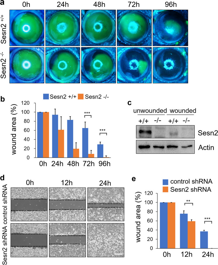
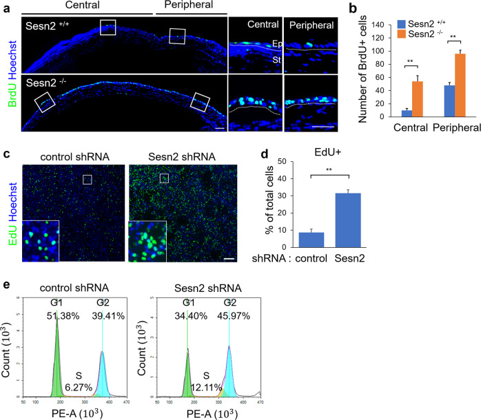
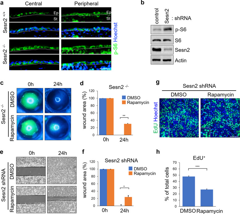
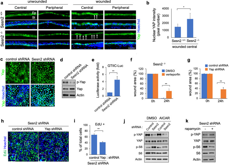
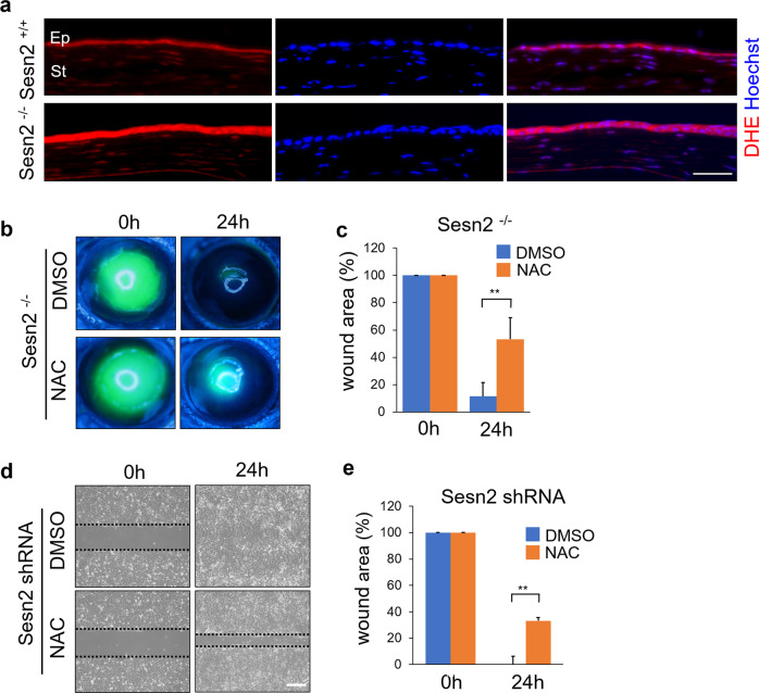
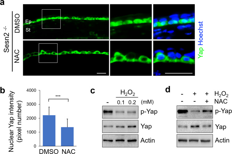
Corneal wound healing is essential for the maintenance of corneal integrity and transparency and involves a series of physiological processes that depend on the proliferation of epithelial cells. However, the molecular mechanisms that control corneal epithelial cell proliferation are poorly understood. Here, we show that Sestrin2, a stress-inducible protein, is downregulated in the corneal epithelium during wound healing and that the proliferation of epithelial basal cells is enhanced in Sestrin2-deficient mice. We also show that YAP, a major downstream effector of the Hippo signaling pathway, regulates cell proliferation during corneal epithelial wound repair and that Sestrin2 suppresses its activity. Moreover, increased levels of reactive oxygen species in the Sestrin2-deficient corneal epithelium promote the nuclear localization and dephosphorylation of YAP, activating it to enhance the proliferation of corneal epithelial cells. These results reveal that Sestrin2 is a negative regulator of YAP, which regulates the proliferative capacity of basal epithelial cells, and may serve as a potential therapeutic target for corneal epithelial damage.


Conflict of interest statement

The authors declare that they have no conflict of interest.

Figures

Fig. 1
Fig. 1. Sesn2 deficiency enhances corneal wound healing.
a Representative photographs of the fluorescein-stained corneas of Sesn2−/− and Sesn2+/+ mice at 0, 24, 48, 72, and 96 h after injury using a 2.5 mm punch. The fluorescein-stained area represents the corneal epithelial wound area. By 96 h, the corneal wound area in Sesn2−/− mice was healed, whereas that of Sesn2+/+ mice remained open. b Quantitative analysis of the corneal wound area of Sesn2−/− and +/+ mice. A total of 12 eyes from Sesn2−/− and Sesn2+/+ mice were used to measure the epithelial wound area size. The wound area was significantly smaller in Sesn2−/− mice than in Sesn2+/+ mice. c Corneal tissues from Sesn2−/− and Sesn2+/+ mice were lysed 48 h after injury and subjected to western blotting with Sesn2 and actin antibodies. d In vitro wound healing assays of hCET cells expressing Sesn2 shRNA and control shRNA. hCET cells expressing Sesn2 shRNA or control shRNA were seeded on both sides of a wound chamber and allowed to attach for 12 h. The chamber was removed, and the wound areas were photographed immediately at 0, 12, and 24 h. Dotted lines indicate wound borders at the beginning of the assay. e Quantitative analysis of the wound areas of hCET cells expressing Sesn2 shRNA and control shRNA at 0, 12, and 24 h. The rate of wound closure in hCET cells expressing Sesn2 shRNA was significantly higher than in hCET cells expressing control shRNA. Error bars represent the means ± SD of three independent experiments. Two-tailed Student’s t-test (**p < 0.01, ***p < 0.001). Scale bar, 300 µm.
Fig. 2
Fig. 2. Sesn2 deficiency promotes corneal epithelial cell proliferation.
a BrdU was injected into Sesn2−/− and Sesn2+/+ mice after injury, and the mice were sacrificed 48 h. Corneal sections were stained using an anti-BrdU antibody (green). Hoechst dye was used as a counterstain (blue). BrdU-positive cells were observed in the central and peripheral regions of corneal epithelial layers and were primarily detected in Sesn2−/− mice. Dotted lines indicate the basement membrane. b Quantitative analysis of BrdU-positive cells in the corneal epithelium of Sesn2−/− and Sesn2+/+ mice after injury. c EdU incorporation assay of hCET cells expressing Sesn2 shRNA or control shRNA. Cells were incubated with 10 µM EdU for 4 h. d Percentage of EdU-positive cells. The number of EdU-positive Sesn2-deficient hCET cells was significantly increased. e Distribution of cells in different cell cycle phases. The proportion of Sesn2-deficient hCET cells in the S and G2 phases of the cell cycle was higher than that of control cells. Error bars represent the means ± SD of three independent experiments. Two-tailed Student’s t-test (**p < 0.01). Scale bars, 50 µm in (a); 100 µm in (c).
Fig. 3
Fig. 3. Sesn2 deficiency increases mTOR signaling activity.
a Sections of injured corneas from Sesn2−/− and Sesn2+/+ mice were immunostained using an anti-phospho-S6 antibody. Phospho-S6 expression was increased in the corneal epithelium of Sesn2−/− mice compared to that in the corneal epithelium of Sesn2+/+ mice. Dotted lines indicate the basement membrane. b hCET cells expressing Sesn2 shRNA or control shRNA were subjected to western blotting with phospho-S6, S6, Sesn2, and actin antibodies. c In vivo corneal epithelial wound healing in Sesn2−/− mice after injury using a 2 mm punch. Each cornea was treated with 100 nM rapamycin or DMSO after injury. At 24 h, the wound area of rapamycin-treated Sesn2−/− mice remained open, whereas that of DMSO-treated Sesn2−/− mice had healed. d Quantitative analysis of the wound area of rapamycin- and DMSO-treated Sesn2−/− mice. Wound closure in rapamycin-treated Sesn2−/− mice was significantly delayed compared to that in DMSO-treated Sesn2−/− mice. e In vitro wound healing assay of hCET cells expressing Sesn2 shRNA treated with 100 nM rapamycin or DMSO. f Quantitative analysis of wound area showing that the rate of wound closure in rapamycin-treated cells was significantly delayed compared to that in DMSO-treated cells. g EdU incorporation assay of Sesn2-deficient hCET cells treated with rapamycin or DMSO. h The number of EdU-positive cells was decreased in rapamycin-treated cultures compared to DMSO-treated cultures. Error bars represent the mean ± SD of three independent experiments. Two-tailed Student’s t-test (*p < 0.05, **p < 0.01, ***p < 0.001). Scale bars, 50 µm in (a, g); 300 µm in (e).
Fig. 4
Fig. 4. Sesn2 negatively regulates YAP activity.
a Sections of injured and normal corneas from Sesn2−/− and Sesn2+/+ mice were immunostained using an anti-YAP antibody. YAP is expressed in the basal layer of the peripheral and central corneal epithelium. White arrows indicate the cells showing YAP nuclear localization. b Fluorescence intensity of nuclear YAP in the epithelial cells of the central cornea from Sesn2−/− and Sesn2+/+ mice. c Sesn2 deficiency increased YAP nuclear localization. hCET cells expressing Sesn2 shRNA or control shRNA were immunostained using an anti-YAP antibody. Hoechst dye was used as a counterstain. d Sesn2 deficiency increased YAP activity. hCET cells expressing Sesn2 shRNA or control shRNA were subjected to western blotting with antibodies against phospho-YAP, YAP, and actin. e hCET cells expressing Sesn2 shRNA or control shRNA were transfected with the GTIIC-Lux reporter. Luciferase activity in each sample was measured as described in the Materials and Methods section. f Quantitative analysis of corneal epithelial wound healing in Sesn2−/− mice after injury using a 2 mm punch. Each cornea was treated with 20 µM verteporfin or DMSO after injury. The wound area was significantly smaller in DMSO-treated corneas than in verteporfin-treated corneas. g Quantitative analysis of the in vitro wound healing of Sesn2-deficient hCET cells expressing YAP shRNA or control shRNA. Wound closure in hCET cells expressing YAP shRNA was significantly delayed compared to that in hCET cells expressing control shRNA. h EdU incorporation assay of Sesn2-deficient hCET cells expressing YAP shRNA or control shRNA. i Percentage of EdU-positive cells. YAP deficiency decreased the proliferation of corneal epithelial cells. j hCET cells expressing Sesn2 shRNA or control shRNA were treated with 0.5 mM AICAR or DMSO and subjected to western blotting with phospho-YAP, YAP, phospho-S6, S6, and actin antibodies. k Western blot analysis of cell lysates from Sesn2 shRNA-expressing hCET cells treated with rapamycin or DMSO. Rapamycin treatment did not affect phospho-Yap expression. Error bars represent the means ± SD of three or four independent experiments. Two-tailed Student’s t-test (*p < 0.05, **p < 0.01). Scale bars, 50 µm in (c, h); 100 µm in (a).
Fig. 5
Fig. 5. ROS levels increase in Sesn2-deficient corneal epithelium, and the inhibition of ROS suppresses corneal wound healing.
a Detection of ROS by DHE staining in the sections of the injured corneas from Sesn2−/− and Sesn2+/+ mice. DHE fluorescence intensity in the corneas of Sesn2−/− mice was significantly higher than that in the corneas of Sesn2+/+ mice. b In vivo corneal epithelial wound healing of Sesn2−/− mice after injury using a 2 mm punch. Each cornea was treated with 2 mM NAC or DMSO after injury. c Quantitative analysis of the corneal wound area of Sesn2−/− mice treated with NAC or DMSO. Inhibition of ROS suppressed corneal wound healing. d In vitro wound healing assay of hCET cells expressing Sesn2 shRNA treated with 2 mM NAC or DMSO. e Wound closure in NAC-treated hCET cells was delayed than that in DMSO-treated cells. Error bars represent the means ± SD of three independent experiments. Two-tailed Student’s t-test (**p < 0.01). Scale bar, 50 µm in (a); 300 µm in (d).
Fig. 6
Fig. 6. YAP is activated by increased ROS production.
a Sections of injured corneas from Sesn2−/− mice treated with 2 mM NAC or DMSO were immunostained using an anti-YAP antibody. Treatment with NAC decreased YAP nuclear localization. b Fluorescence intensity of nuclear YAP in corneal epithelial cells treated with NAC or DMSO. The nuclear YAP fluorescence intensity of NAC-treated corneal epithelial cells was significantly lower than that of DMSO-treated cells. c Treatment with H2O2 increased YAP activity. hCET cells were treated with 0.1 or 0.2 mM H2O2 for 2 h. Cell lysates were subjected to western blotting with phospho-YAP, YAP, and actin antibodies. d Treatment with NAC suppressed YAP activity. hCET cells were treated with 0.2 mM H2O2 or 2 mM NAC and subjected to western blotting with phospho-YAP, YAP, and actin antibodies. Error bars represent the means ± SD of three independent experiments. Two-tailed Student’s t-test (***p < 0.001). Scale bar, 50 µm.

Similar articles

Cited by

References

    1. Tseng SCG. Concept and application of limbal stem cells. Eye. 1989;3:141–157. - PubMed
    1. Sun TT, Lavker RM. Corneal epithelial stem cells: past, present, and future. J. Investig. Dermatol. Symp. Proc. 2004;9:202–207. - PubMed
    1. Ljubimov AV, Saghizadeh M. Progress in corneal wound healing. Prog. Retin. Eye Res. 2015;49:17–45. - PMC - PubMed
    1. Lee JH, Budanov AV, Karin M. Sestrins orchestrate cellular metabolism to attenuate aging. Cell Metab. 2013;18:792–801. - PMC - PubMed
    1. Budanov AV, Sablina AA, Feinstein E, Koonin EV, Chumakov PM. Regeneration of peroxiredoxins by p53-regulated Sestrins, homologs of bacterial AhpD. Science. 2004;304:596–600. - PubMed

Publication types