

Pituispheres Contain Genetic Variants Characteristic to Pituitary Adenoma Tumor Tissue
- PMID: 32528411
- PMCID: PMC7256168
- DOI: 10.3389/fendo.2020.00313
Pituispheres Contain Genetic Variants Characteristic to Pituitary Adenoma Tumor Tissue
Abstract
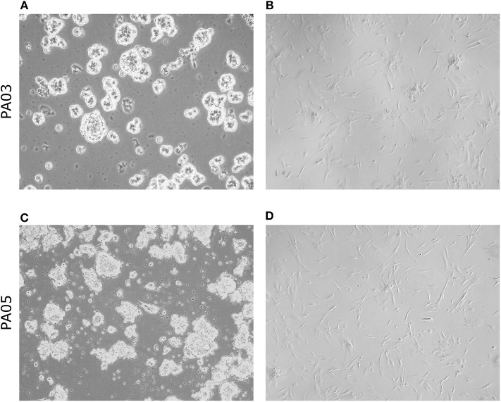
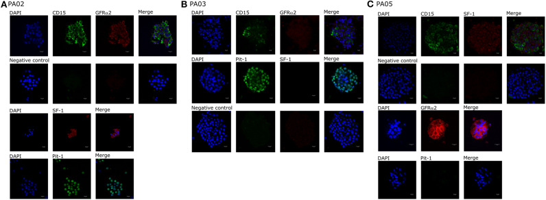
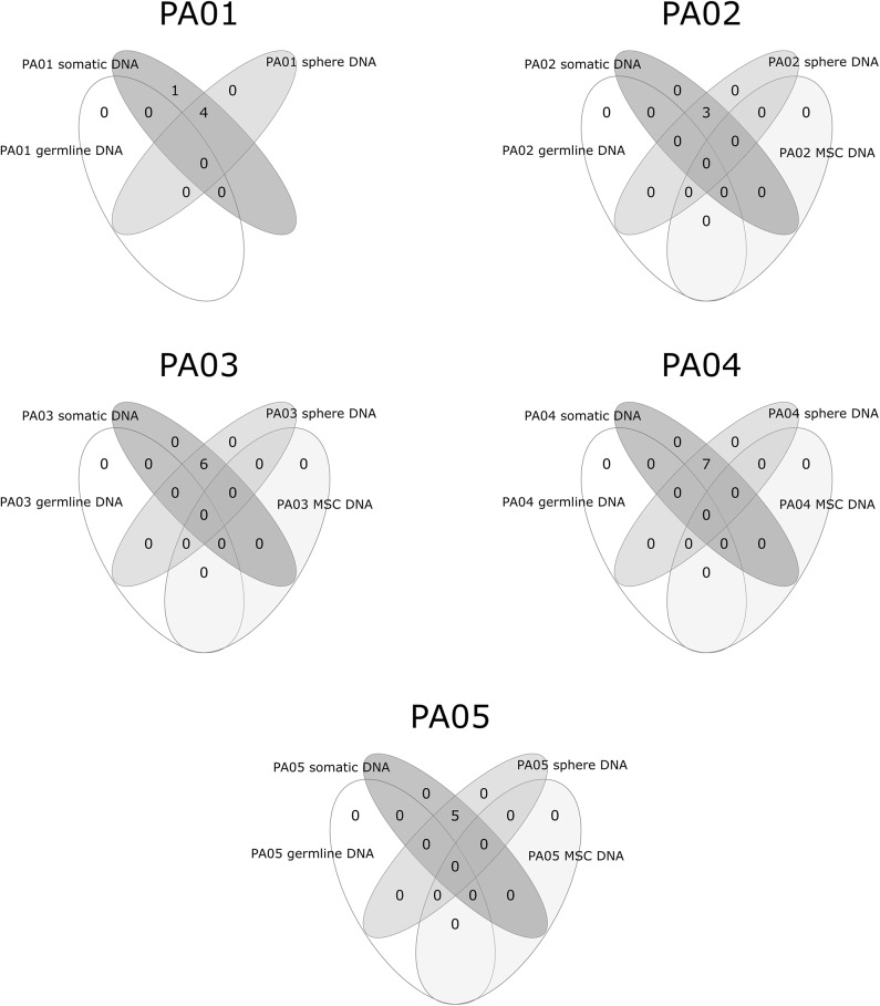
The most common type of pituitary neoplasms is benign pituitary adenoma (PA). Clinically significant PAs affect around 0.1% of the population. Currently, there is no established human PA cell culture available and when PA tumor cells are cultured they form two distinct types depending on culturing conditions either free-floating aggregates also known as pituispheres or cells adhering to the surface of cell plates and displaying mesenchymal stem-like properties. The aim of this study was to trace the origin of sphere-forming and adherent pituitary cell cultures and characterize the potential use of these surgery derived cell lines as PA model. We carried out a paired-end exome sequencing of patients' tumor and germline DNA using Illumina NextSeq followed by characterization of corresponding PA cell cultures. Variation analysis revealed a low amount of somatic mutations (mean = 5.2, range 3-7) in exomes of PAs. Somatic mutations of the primary surgery material can be detected in the exomes of respective pituispheres, but not in exomes of respective mesenchymal stem-like cells. For the first time, we show that the genome of pituispheres represents genome of PA while mesenchymal stem cells derived from the PA tissue do not contain mutations characteristic to PA in their genome, therefore, most likely representing normal cells of pituitary or surrounding tissues. This finding indicates that pituispheres can be used as a human model of PA cells, but combination of cell culturing techniques and NGS needs to be employed to adjust for disability to propagate spheres in culturing conditions.
Keywords: pituispheres; pituitary adenoma; pituitary adenoma cultures; tumor sequencing; whole exome sequencing.
Copyright © 2020 Peculis, Mandrika, Petrovska, Dortane, Megnis, Nazarovs, Balcere, Stukens, Konrade, Pirags, Klovins and Rovite.
Figures

Similar articles
-
A pilot genome-scale profiling of DNA methylation in sporadic pituitary macroadenomas: association with tumor invasion and histopathological subtype.PLoS One. 2014 Apr 29;9(4):e96178. doi: 10.1371/journal.pone.0096178. eCollection 2014. PLoS One. 2014. PMID: 24781529 Free PMC article.
-
N-myc downstream-regulated gene 2 (NDRG2) promoter methylation and expression in pituitary adenoma.Diagn Pathol. 2017 Apr 8;12(1):33. doi: 10.1186/s13000-017-0622-7. Diagn Pathol. 2017. PMID: 28390436 Free PMC article.
-
Pituitary tumors contain a side population with tumor stem cell-associated characteristics.Endocr Relat Cancer. 2015 Aug;22(4):481-504. doi: 10.1530/ERC-14-0546. Epub 2015 Apr 28. Endocr Relat Cancer. 2015. PMID: 25921430
-
HEREDITARY PITUITARY TUMOR SYNDROMES: GENETIC AND CLINICAL ASPECTS.Rev Invest Clin. 2020;72(1):8-18. doi: 10.24875/RIC.19003186. Rev Invest Clin. 2020. PMID: 32132734 Review.
-
MicroRNAs and Target Genes in Pituitary Adenomas.Horm Metab Res. 2018 Mar;50(3):179-192. doi: 10.1055/s-0043-123763. Epub 2018 Jan 19. Horm Metab Res. 2018. PMID: 29351706 Review.
Cited by
-
Clinical Biology of the Pituitary Adenoma.Endocr Rev. 2022 Nov 25;43(6):1003-1037. doi: 10.1210/endrev/bnac010. Endocr Rev. 2022. PMID: 35395078 Free PMC article. Review.
-
Identification and gene expression profiling of human gonadotrophic pituitary adenoma stem cells.Acta Neuropathol Commun. 2023 Feb 7;11(1):24. doi: 10.1186/s40478-023-01517-w. Acta Neuropathol Commun. 2023. PMID: 36750863 Free PMC article.
-
Architects of Pituitary Tumour Growth.Front Endocrinol (Lausanne). 2022 Jun 28;13:924942. doi: 10.3389/fendo.2022.924942. eCollection 2022. Front Endocrinol (Lausanne). 2022. PMID: 35837315 Free PMC article. Review.
-
Comprehensive transcriptomic analysis identifies three distinct subtypes of pituitary adenomas: insights into tumor behavior, prognosis, and stem cell characteristics.J Transl Med. 2024 Oct 3;22(1):892. doi: 10.1186/s12967-024-05702-w. J Transl Med. 2024. PMID: 39363281 Free PMC article.
-
Large Scale Molecular Studies of Pituitary Neuroendocrine Tumors: Novel Markers, Mechanisms and Translational Perspectives.Cancers (Basel). 2021 Mar 19;13(6):1395. doi: 10.3390/cancers13061395. Cancers (Basel). 2021. PMID: 33808624 Free PMC article. Review.
References
-
- Avinash Kumar K. MRI measurement of normal pituitary size using volumetric imaging in Scottish patients. Curr Trends Clin Med Imaging. (2017) 1:1–5. 10.19080/CTCMI.2017.01.555563 - DOI
Publication types
MeSH terms
Substances
LinkOut - more resources
Full Text Sources
Medical