The Keloid Disorder: Heterogeneity, Histopathology, Mechanisms and Models
- PMID: 32528951
- PMCID: PMC7264387
- DOI: 10.3389/fcell.2020.00360
The Keloid Disorder: Heterogeneity, Histopathology, Mechanisms and Models
Abstract
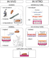
Keloids constitute an abnormal fibroproliferative wound healing response in which raised scar tissue grows excessively and invasively beyond the original wound borders. This review provides a comprehensive overview of several important themes in keloid research: namely keloid histopathology, heterogeneity, pathogenesis, and model systems. Although keloidal collagen versus nodules and α-SMA-immunoreactivity have been considered pathognomonic for keloids versus hypertrophic scars, conflicting results have been reported which will be discussed together with other histopathological keloid characteristics. Importantly, histopathological keloid abnormalities are also present in the keloid epidermis. Heterogeneity between and within keloids exists which is often not considered when interpreting results and may explain discrepancies between studies. At least two distinct keloid phenotypes exist, the superficial-spreading/flat keloids and the bulging/raised keloids. Within keloids, the periphery is often seen as the actively growing margin compared to the more quiescent center, although the opposite has also been reported. Interestingly, the normal skin directly surrounding keloids also shows partial keloid characteristics. Keloids are most likely to occur after an inciting stimulus such as (minor and disproportionate) dermal injury or an inflammatory process (environmental factors) at a keloid-prone anatomical site (topological factors) in a genetically predisposed individual (patient-related factors). The specific cellular abnormalities these various patient, topological and environmental factors generate to ultimately result in keloid scar formation are discussed. Existing keloid models can largely be divided into in vivo and in vitro systems including a number of subdivisions: human/animal, explant/culture, homotypic/heterotypic culture, direct/indirect co-culture, and 3D/monolayer culture. As skin physiology, immunology and wound healing is markedly different in animals and since keloids are exclusive to humans, there is a need for relevant human in vitro models. Of these, the direct co-culture systems that generate full thickness keloid equivalents appear the most promising and will be key to further advance keloid research on its pathogenesis and thereby ultimately advance keloid treatment. Finally, the recent change in keloid nomenclature will be discussed, which has moved away from identifying keloids solely as abnormal scars with a purely cosmetic association toward understanding keloids for the fibroproliferative disorder that they are.
Keywords: cicatrix, hypertrophic; keloid; keloid anatomy and histology; keloid etiology; keloid heterogeneity; keloid model; keloid pathology; keloid phenotype.
Copyright © 2020 Limandjaja, Niessen, Scheper and Gibbs.
Figures


References
-
- Agbenorku P. T., Kus H., Myczkowski T. (1995). The keloid triad hypothesis (KTH): a basis for keloid etiopathogenesis and clues for prevention. Eur. J. Plast. Surg. 18 301–304.
Publication types
LinkOut - more resources
Full Text Sources
Research Materials

