

Staphyloma-related chorioretinal folds
- PMID: 32529117
- PMCID: PMC7281792
- DOI: 10.1016/j.ajoc.2020.100747
Staphyloma-related chorioretinal folds
Abstract
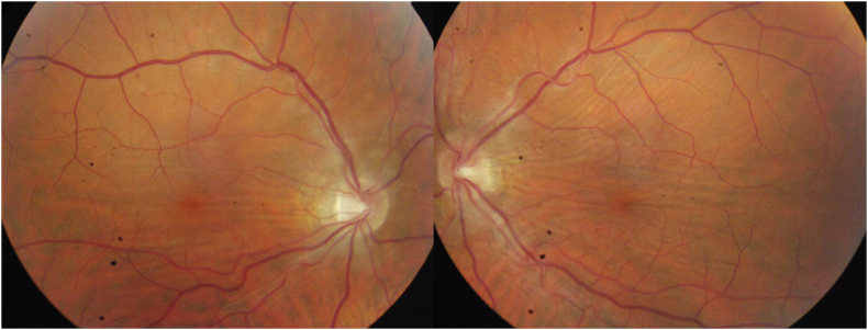
Purpose: To report a case of bilateral idiopathic chorioretinal folds that seemed to be related to an atypical staphyloma.
Observations: A 49-year old man without medical history consulted for slight vision loss and metamorphopsia in the left eye. The ophthalmologic examination revealed moderate myopia and bilateral chorioretinal folds in the posterior pole, confirmed by multimodal imaging. Orbital and systemic examinations ruled out all the known etiologies of chorioretinal folds. 3-D optical coherence tomography and B-scan suggested that the folds were related to an atypical staphyloma that developed in the temporal part of the fundus, while sparing the peripapillary area. The peripapillary area, spared by the staphyloma process, appeared as a "dome-shaped disc" compared to the staphylomatous area.
Conclusion and importance: This case suggests that myopic patients with unusual staphyloma located outside the peripapillary area could develop chorioretinal folds.
Keywords: Chorioretinal folds; Dome-shaped disc; Multimodal imaging; Myopia; Staphyloma.
© 2020 The Authors.
Conflict of interest statement
The following authors have no financial disclosures related to this study: AGA, CL, AG, TG, SYC. Financial disclosures not related to the study: AGA is consultant for Allergan, Bayer and Novartis. AG is consultant for Novartis and Thrombogenics. TG is consultant for Novartis. SYC is consultant for Allergan, Bayer, Novartis, Roche, Thea and Tilak.
Figures


References
-
- Leahey A.B., Brucker A.J., Wyszynski R.E., Shaman P. Chorioretinal folds. A comparison of unilateral and bilateral cases. Arch Ophthalmol. 1993;111(3):357–359. - PubMed
-
- Gass J.D. Radial chorioretinal folds. A sign of choroidal neovascularization. Arch Ophthalmol. 1981;99(6):1016–1018. - PubMed
-
- Ahmadi A.J., Pirinjian G.E., Sires B.S. Optic neuropathy and macular chorioretinal folds caused by orbital cherubism. Arch Ophthalmol. 2003;121(4):570–573. - PubMed
-
- Yeung L., Lai C.C., Chen T.L., Wu W.C. Chorioretinal folds associated with a meningioma. Chang Gung Med J. 2005;28(8):575–580. - PubMed
-
- Hyvärinen L., Walsh F.B. Benign chorioretinal folds. Am J Ophthalmol. 1970;70(1):14–17. - PubMed
Publication types
LinkOut - more resources
Full Text Sources
