. 2020 Aug;140(2):183-208.
doi: 10.1007/s00401-020-02174-2. Epub 2020 Jun 11.

HIF-1α is involved in blood-brain barrier dysfunction and paracellular migration of bacteria in pneumococcal meningitis

Affiliations

HIF-1α is involved in blood-brain barrier dysfunction and paracellular migration of bacteria in pneumococcal meningitis

Gayatri Devraj et al. Acta Neuropathol. 2020 Aug.

Abstract

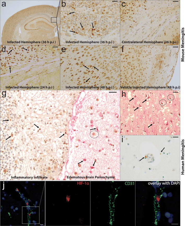
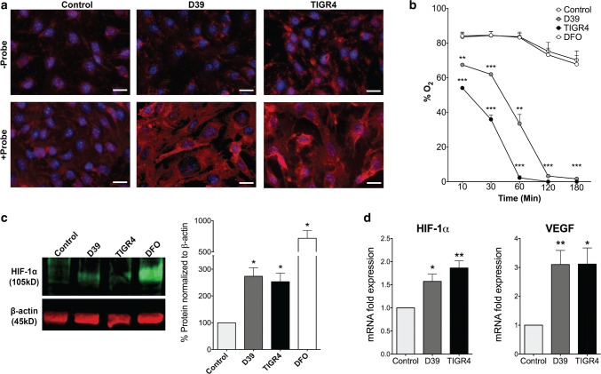
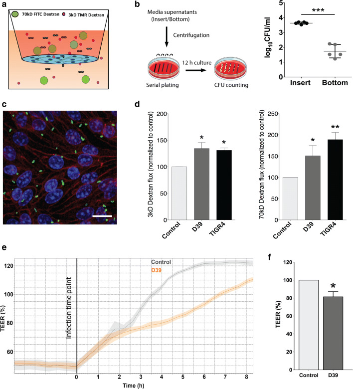
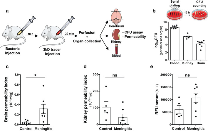
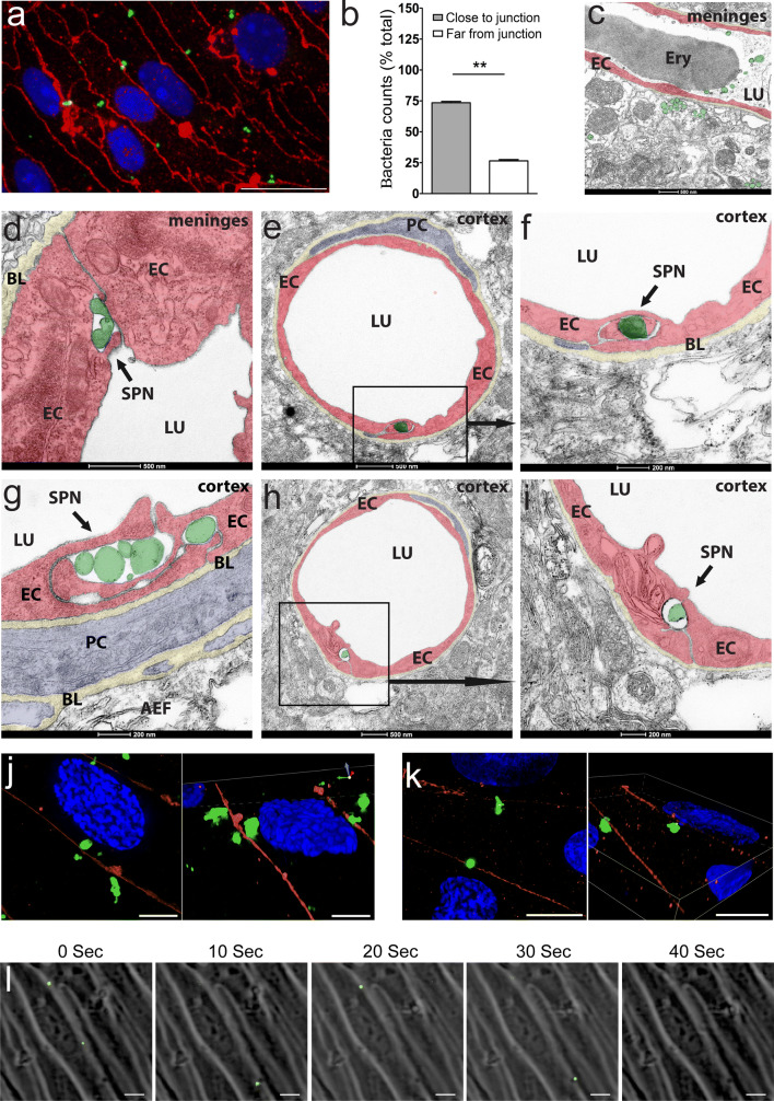
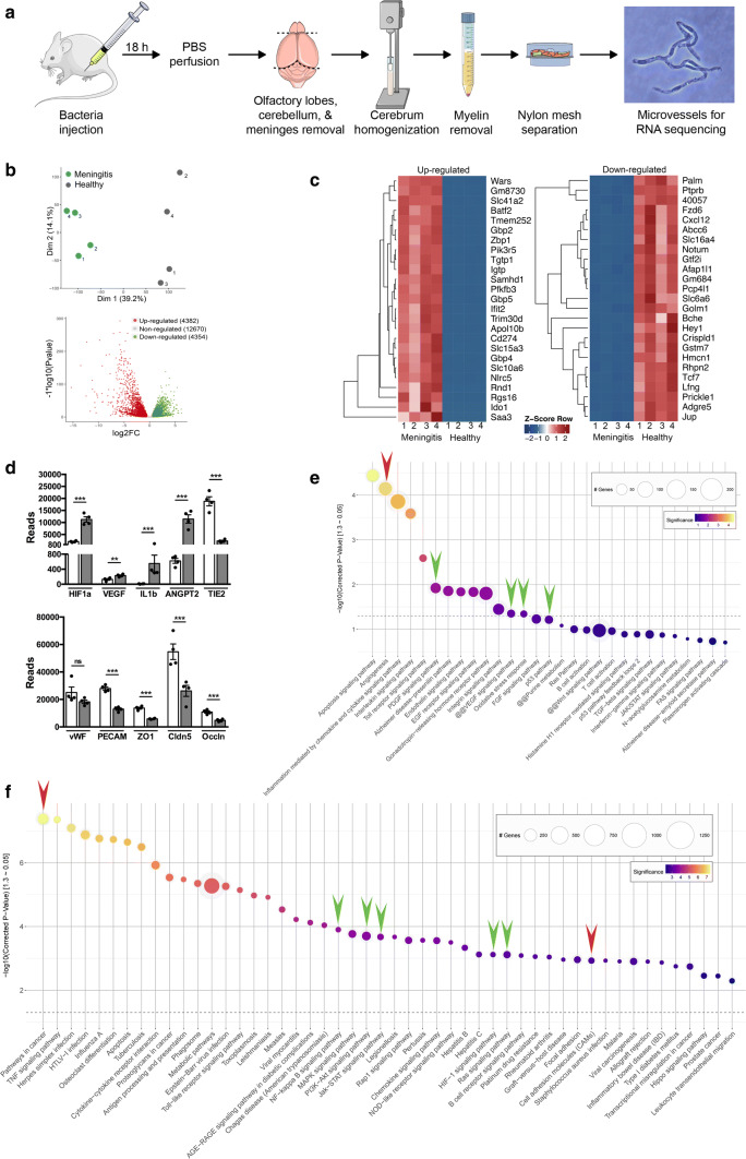
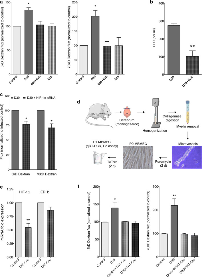
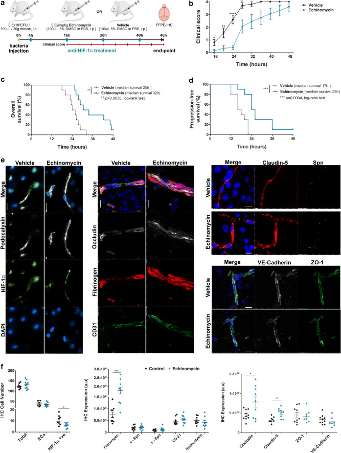
Bacterial meningitis is a deadly disease most commonly caused by Streptococcus pneumoniae, leading to severe neurological sequelae including cerebral edema, seizures, stroke, and mortality when untreated. Meningitis is initiated by the transfer of S. pneumoniae from blood to the brain across the blood-cerebrospinal fluid barrier or the blood-brain barrier (BBB). The underlying mechanisms are still poorly understood. Current treatment strategies include adjuvant dexamethasone for inflammation and cerebral edema, followed by antibiotics. The success of dexamethasone is however inconclusive, necessitating new therapies for controlling edema, the primary reason for neurological complications. Since we have previously shown a general activation of hypoxia inducible factor (HIF-1α) in bacterial infections, we hypothesized that HIF-1α, via induction of vascular endothelial growth factor (VEGF) is involved in transmigration of pathogens across the BBB. In human, murine meningitis brain samples, HIF-1α activation was observed by immunohistochemistry. S. pneumoniae infection in brain endothelial cells (EC) resulted in in vitro upregulation of HIF-1α/VEGF (Western blotting/qRT-PCR) associated with increased paracellular permeability (fluorometry, impedance measurements). This was supported by bacterial localization at cell-cell junctions in vitro and in vivo in brain ECs from mouse and humans (confocal, super-resolution, electron microscopy, live-cell imaging). Hematogenously infected mice showed increased permeability, S. pneumoniae deposition in the brain, along with upregulation of genes in the HIF-1α/VEGF pathway (RNA sequencing of brain microvessels). Inhibition of HIF-1α with echinomycin, siRNA in bEnd5 cells or using primary brain ECs from HIF-1α knock-out mice revealed reduced endothelial permeability and transmigration of S. pneumoniae. Therapeutic rescue using the HIF-1α inhibitor echinomycin resulted in increased survival and improvement of BBB function in S. pneumoniae-infected mice. We thus demonstrate paracellular migration of bacteria across BBB and a critical role for HIF-1α/VEGF therein and hence propose targeting this pathway to prevent BBB dysfunction and ensuing brain damage in infections.

Keywords: Blood–brain barrier (BBB); Dexamethasone; Endothelium; HIF-1α; Meningitis; Paracellular transmigration; Permeability; S. pneumoniae; VEGF.


Conflict of interest statement

The authors declare no conflict of interest.

Figures

Fig. 1
Fig. 1
Induction of HIF-1α in mouse and human brain tissue samples from pneumococcal meningitis. a Immunohistochemistry of brain tissue 36 h post-S. pneumoniae infection in intracerebrally infected mouse model shows positive HIF-1α in several inflammatory and neural cells. b Zoomed area from a in the cortex region of the infected mouse brain shows significant nuclear HIF-1α (black arrows) indicating its activation along with positive staining in the granulocytic infiltrate. c Non-infected hemisphere of the same mouse at 36 h post infection shows no specific staining for HIF-1α signal. d Positive staining in the cortex region (black arrows) was also observed at 24 h post-intracerebral infection, which was also observed at e 48 h post-infection in the intracisternally infected mice. f Control mice that were intracerebrally injected with 0.9% NaCl did not show any positive staining in the same region. g Brain sections from human pneumococcal meningitis patients double stained for HIF-1α (brown) and GFAP (red) show nuclear staining for HIF-1α (black arrows) in lymphocytes of the inflammatory infiltrate (g, left) and in the edematous brain tissue comprising GFAP positive reactive astrocytes (g, right), where macrophages showed cytoplasmic staining (black circles). h Neurons labeled with MAP2 (red) were also co-stained with HIF-1α (brown) in the cortex but mostly the pyknotic ones and not the vital neurons (black circles). i Positive HIF-1α staining could also be observed in vessel associated cells in the cortex region. j Activated HIF-1α indicated by nuclear staining (red) could be observed in brains ECs from pneumococcal meningitis patients by immunofluorescence staining using CD31 as an endothelial marker. Scale bar bf is 20 µm, gi is 25 µm, j left most image 20 µm and the zoomed images 10 µm. Mouse brain stainings are representative of three mice each at 24, 36, 48 h post-infection and two control mice at 48 h post- vehicle injection. Human pneumococcal meningitis stainings are representative of 4 cases outlined in Table 1
Fig. 2
Fig. 2
Hypoxia and HIF-1α/VEGF induction upon S. pneumoniae infection in brain ECs. a Control and S. pneumoniae infected bEnd5 cells treated with pimonidazole hydrochloride (Hypoxyprobe 200 mM) and visualized for cellular hypoxia by red 549-linked mouse anti-pimonidazole monoclonal antibodies. D39 and TIGR4 strains infected cells show significantly high number of hypoxic cells compared to control cells which is however not present in the top row without the probe indicating specificity of the staining (scale bar 10 µm). b Oxygen levels (%) were quantified in the medium of control and S. pneumoniae infected bEnd5 cells using the SensorDish® Reader (SDR) for non-invasive measurement of O2 levels through the transparent well bottom. O2 concentration (pO2% air saturation) was obtained at 60-s intervals and average values at several different time points shown indicate a significant reduction in O2 levels within minutes post-infection and to hypoxic levels by 2 h (N = 3, 2-tailed paired t test compared to control for each condition at the corresponding time point, mean ± SEM, **p < 0.01, ***p < 0.001), which was however not present either in control or deferoxamine (DFO, 200 µM), an iron chelator that induces HIF-1α independent of hypoxia. c Control and S. pneumoniae infected bEnd5 cells were analyzed for HIF-1α protein induction by Western blotting. The result shows a significant increase in HIF-1α levels in both D39 and TIGR4 infected cells compared to control conditions when the band pixel intensity is quantified by normalizing to the housekeeping protein β-actin band. DFO served as a positive control for HIF-1α induction (N = 4, *p < 0.05, 2-tailed paired t test compared to control for each condition). d Quantitative RT-PCR expression analysis (by 2−∆∆Ct method) of control and infected bEnd5 cells shows upregulation of HIF-1α and its transcriptional target gene VEGF upon a S. pneumoniae infection. Expression was normalized to ribosomal protein, large, P0 (RPLP0) that served as a housekeeping gene (N = 6, *p < 0.05, **p < 0.01, 2-tailed paired t test compared to control)
Fig. 3
Fig. 3
Bacterial transmigration and permeability upon S. pneumoniae infection in brain ECs. a The scheme depicts the in vitro model of the BBB for meningitis infection where bEnd5 cells were cultured in 24-well PET transwells (105 cells/cm2) and infected with S. pneumoniae (D39 and TIGR4 strain; MOI 50) along with the addition of labeled dextrans to the apical chamber (5 μM each of 3 kD TMR, and 70 kD FITC dextran). b Media supernatant from infected and control cells from the insert and the bottom well at 4 h post-infection were collected and centrifuged for plating and culture on blood agar plate. The colony forming unit (CFU) assay indicates transmigration of approximately 1% of the bacteria added to the insert (N = 5, ***p < 0.001, 2-tailed paired t test compared to control). c Confocal microscopy analysis of transwell inserts to confirm the presence of bacteria indicated predominant localization of the bacteria (green) at the cell–cell borders with VE-cadherin (red) serving as EC junction marker (representative from N = 3 experiments; scale bar 10 μm). d Permeability analysis of bEnd5 cells 4 h post-infection performed with low (3 kD) and high molecular weight (70 kD) dextrans shows a significant increase in dextran permeability for both sizes, confirming the breakdown of endothelial barrier due to infection (N = 5, *p < 0.05, **p < 0.01, 2-tailed paired t-test compared to control). e Representative graph for continuous transendothelial electrical resistance (TEER) values of S. pneumoniae (D39) infected bEnd5 cells on inserts using the cellZscope® system setting the values of the control group between 3–4 h post-infection to 100%. The graph shows reduced TEER values starting 3 h post-infection that persists for several hours indicating increase in paracellular permeability. The increase in the TEER values of the control group from the plateau at 0 h reflects the changes due to electrode handling and media composition. f Quantification at 4 h shows a significant reduction in TEER values in infected cells compared to control (N = 4, *p < 0.05, 2-tailed paired t test)
Fig. 4
Fig. 4
Extravasation of S. pneumoniae and neurovascular permeability in vivo in a hematogenous meningitis model. a Setup showing the murine infection model with intraperitoneal injection of 0.5 × 105 bacteria (in 100 μl PBS, D39 strain). 18 h post-infection mice were injected with a 3kD TMR tracer (i.p), circulated for 20 min followed by anesthesia, transcardial PBS perfusion and collection of blood, brain, and kidney. b Homogenized organs or blood were plated and cultured overnight to obtain the bacterial counts using the CFU assay. High CFU values in blood indicated the hematogenous presence of bacteria with extravasation of bacteria evident in the brain (0.01%). The bacterial counts in the kidney were 100-fold higher than brain reflecting higher permeability of the kidney vasculature. Healthy control mice did not have any bacterial growth on the plates. c Homogenized hemi-brain, kidney and blood were utilized to obtain dextran permeability by measuring fluorescence intensity on a microplate reader. A significant elevation in vascular permeability of infected mice was observed compared to healthy controls in the brain indicating blood–brain barrier breakdown. d Vascular permeability of kidney was not altered upon infection. e Serum fluorescence values (arbitrary units—a.u.) indicate equivalent tracer absorption between healthy and infected mice. (N = 5–7/group, *p < 0.05, 2-tailed unpaired t test)
Fig. 5
Fig. 5
Junctional localization of S. pneumoniae at the BBB in vitro and in vivo. a Primary mouse brain ECs (MBMEC) were infected with S. pneumoniae and subjected to immunofluorescence staining for claudin-5 to mark endothelial junctions (red) and co-stained with an anti-pneumococcal antibody (green) to label S. pneumoniae. Confocal fluorescence microscopy of EC monolayers indicated a predominant localization of bacteria at the cell–cell borders co-localizing with claudin-5, an endothelial tight junctions marker as shown by b quantification that indicated a significantly higher number of bacteria close to the junctions. N = 3 independent preparations of MBMEC from 2–3 mice each time. Two independent wells were counted for each set comprising approximately 25 cells/field at 60X magnification (scale bar: 20 μm). Bacteria within a distance of 1 bacterium equivalent size from the junction were considered close to the junction. Data presented as mean ± SEM, **p < 0.01, 2-tailed paired t test. c–i Transmission electron microscopy (TEM) analysis was performed on brain sections from hematogenously infected mice post-perfusion with PBS/PFA and fixation in PFA/glutaraldehyde. S. pneumoniae either directly or in protective membrane bound vesicles were localized at the endothelial tight junctions both in meninges and cortex in several mice analyzed (representative images from N = 6 mice subjected to TEM). Artificial coloration was performed for better visualization and to highlight the localization of S. pneumoniae. SPN-S. pneumoniae, EC-endothelial cell, PC-pericyte, LU-lumen, BL-basal lamina, AEF-astrocytic endfeet, ERY-erythrocyte. j–k Primary human brain ECs (HBMEC) were also infected with S. pneumoniae and stained with anti-pneumococcal antibody and claudin-5 and subjected to super resolution microscopy using Nikon structured illumination microscopy (N-SIM). In 2 different preparations of HBMEC, the localization of bacteria was primarily at the junctions with super resolution images demonstrating engagement of S. pneumoniae with the endothelial junctions (Figures to the right both in j, k). Scale bar: 5 μm. l Primary mouse brain ECs (MBMEC) were infected with GFP-labeled S. pneumoniae strain (MOI 10) 2 days post-isolation and subjected to live-cell imaging. MBMECs as well as bacteria were imaged in brightfield and GFP fluorescence channel every 10 s starting 1 h post-infection for a total of 2 h. Five time-lapse images (10-s interval) capture transmigration of few bacteria across cell–cell borders (whites lines), which demonstrate paracellular route for transmigration of S. pneumoniae across brain endothelial cells. Representative images from 1 preparation from N = 3 MBMECs preparations (1 animal/set) infected with GFP-labeled D39 strain. Scale bar: 10 μm.
Fig. 6
Fig. 6
Regulation of HIF-1α/VEGF signaling by RNAseq analysis of brain microvessels from S. pneumoniae infected mice. a Schematic of BBB microvessels isolation for RNAseq analysis showing intraperitoneal injection of bacteria followed by anesthesia and transcardial perfusion 18 h post-infection. Extracted brains were cleared of meninges, olfactory lobes, and cerebellum and homogenized followed by myelin removal (density centrifugation). The microvessels were separated by filtering through 100 μm nylon mesh and collected from the top of 40 μm mesh. b The PCA plot shows clear clustering of healthy and meningitis microvessels with the volcano plot showing close to 9000 differentially expressed genes (DEG). c Heatmap analysis shows top 25 upregulated and downregulated genes by Z-score transformation. The data are displayed in a grid where each row represents a gene and each column represents the sample replicate. d Visualization of actual base reads of gene expression by RNAseq profiling shows upregulation of HIF-1α, VEGF and inflammation-related genes such as IL1b and ANGPT2. BBB junction molecules claudin-5, occludin, ZO-1 were downregulated, indicating breakdown of the BBB in this mouse model at the transcriptomic level. e PANTHER pathway analysis of the RNAseq data illustrates the activation of pathways related to angiogenesis (red arrow) and HIF-1α/VEGF signaling, and the ones related to them such as PDGF, p53 pathways (green arrows). f The KEGG pathway enrichment also indicated activation of HIF-1α/VEGF signaling, and related pathways driven by NF-kappa B, PI3-Akt, Jak-STAT, and Ras signaling (green arrows). Pathways related to cancer and vascular remodeling were also activated (red arrows) including those related to inflammation. Dotted line in e, f indicates the cutoff value for significance with color coding for significance level (lighter being more significant) and bubble size reflecting the number of genes in the significant hits (N = 4 samples/group pooling 2 brains per sample)
Fig. 7
Fig. 7
HIF-1α-dependent endothelial permeability and transmigration of S. pneumoniae post-infection. a Treatment of brain ECs (bEnd5) with echinomycin (Ech), an HIF-1α inhibitor, did not lead to higher permeability post-S. pneumoniae infection (D39 strain) for both low (3 kD) and high molecular weight (70kD) dextrans, whereas untreated cells showed increase for both the tracers. Ech alone did not affect permeability when compared to uninfected controls. (mean ± SEM, N = 3, *p < 0.05, 2-tailed paired t test. b Ech treatment also led to significant reduction in the transmigrated bacteria across bEnd5 monolayers, which combined with Fig. 1a indicates HIF-1α dependence of both permeability and paracellular bacterial transmigration (mean ± SEM, N = 3, **p < 0.01, 2-tailed unpaired t test). c Genetic ablation of HIF-1α using siRNA-mediated knockdown in bEnd5 cells also demonstrated a decrease in permeability post-infection compared to scrambled controls (mean ± SEM, N = 4, *p < 0.05, 2-tailed paired t test). d Schematic of primary mouse brain endothelial cells (MBMEC) isolation and culture for permeability analysis post infection. Cerebral hemispheres from HIF-1αflox/flox mice cleared of meninges were homogenized followed by myelin removal and collagenase digestion. Plated microvessels were treated with puromycin to obtain pure EC cultures, followed by treatment with TATcre to induce knockdown of HIF-1α and seeded on to transwell inserts for infection and permeability analysis. e Quantitative RT-PCR confirmed about 50% knockdown of HIF-1α in MBMEC when normalized to RPLP0 (a housekeeping gene), VE-cadherin (CDH5) was not changed (mean ± SEM, N = 5 MBMEC preparations pooling 2–3 mice/set, **p < 0.01, 2-tailed paired t test). f Permeability analysis of MBMEC to both low and high molecular weight dextrans showed an increase post infection with S. pneumoniae (D39) as observed with bEnd5 cells. This effect was abrogated upon HIF-1α knockdown in TAT-cre treated cells demonstrating HIF-1α dependence of permeability post infection also in primary brain endothelium (mean ± SEM, N = 6 MBMEC preparations pooling 2–3 mice/set, *p < 0.05, **p < 0.01, 2-tailed paired t test)
Fig. 8
Fig. 8
Improved survival and blood–brain barrier function post pneumococcal infection in mice by anti-HIF-1α treatment using echinomycin. a Schematic depicting treatment protocol of mice infected with S. pneumoniae (D39). Mice were infected i.p., followed by anti-HIF-1α treatment with echinomycin or just vehicle every 12 h starting 4 h post-infection (blue tick marks). Clinical scores were taken every 4 h starting 16 h post-infection (red line with tick marks) and study terminated at 48 h post-infection and brain tissues collected upon animal death or at the end point when the animals were sacrificed. b Clinical scores indicate improved symptoms in echinomycin-treated mice compared to vehicle-treated animals starting 16 h post-infection that were progression free (below score 1) up to 24 h (mean ± SEM at each time, N = 10/group, ****p < 0.0001, **p < 0.01, *p < 0.05 by 2-tailed unpaired non-parametric Mann–Whitney t test at each time point. c Echinomycin-treated mice showed significantly improved overall survival percentage by Kaplan–Meier analysis with the median survival at 32 h compared to 25 h in vehicle-treated mice (**p < 0.01, log-rank test, N = 10/group). d Kaplan–Meier analysis also indicated a significant improvement in progression-free survival in echinomycin-treated animals (median survival 25 h) with no clinical symptoms up to 24 h (b) compared to the vehicle group (median survival 17 h; ***p < 0.001, log-rank test, N = 10/group). e Immunofluorescence staining for HIF-1α, BBB permeability and junctional markers in the echinomycin and vehicle groups at the survival end point. Left panel shows that echinomycin treatment leads to a reduction in HIF-1α-positive nuclei including in ECs co-stained for podocalyxin, a vascular marker which was unchanged by the treatment. Middle panel displays reduced vascular permeability to fibrinogen in the echinomycin group as indicated by stronger intravascular signal compared to the vehicle-treated mice. Increased expression of tight junction proteins—occludin (middle panel) and claudin-5 (right top panel)—in echinomycin-treated mice indicate improved BBB function. Tight junction-associated ZO-1, adherens junction marker VE-Cadherin, and endothelial cell adhesion molecule CD31 were unchanged (middle, bottom right panel). There was also no difference in S. pneumoniae (Spn) staining (right top panel) between the two groups. Scale bar 10 μm. f Quantification of the staining from e utilizing four images per animal in the cortex region shows a significant reduction in HIF-1α-positive cell number, whereas the total cell number or EC cell number was unchanged (left panel). As observed in e, the middle panel for quantification (arbitrary units—a.u.) of fibrinogen leakage shows significantly increased vascular staining for fibrinogen, supported by significantly increased expression of occludin, claudin-5, whereas other EC junction markers were unchanged. S. pneumoniae numbers in the vessels (v-Spn) or those transmigrated into brain parenchyma (b-Spn) were also unchanged. (mean ± SEM, N = 7–10/group indicated by the corresponding number of dots, ****p < 0.0001, **p < 0.01, *p < 0.05 by 2-tailed unpaired Student’s t test)

References

    1. Antonetti DA, Barber AJ, Hollinger LA, Wolpert EB, Gardner TW. Vascular endothelial growth factor induces rapid phosphorylation of tight junction proteins occludin and zonula occluden 1. A potential mechanism for vascular permeability in diabetic retinopathy and tumors. J Biol Chem. 1999;274:23463–23467. doi: 10.1074/jbc.274.33.23463. - DOI - PubMed
    1. Balconi G, Spagnuolo R, Dejana E. Development of endothelial cell lines from embryonic stem cells: a tool for studying genetically manipulated endothelial cells in vitro. Arterioscler Thromb Vasc Biol. 2000;20:1443–1451. doi: 10.1161/01.atv.20.6.1443. - DOI - PubMed
    1. Barichello T, Collodel A, Generoso JS, Simoes LR, Moreira AP, Ceretta RA, et al. Targets for adjunctive therapy in pneumococcal meningitis. J Neuroimmunol. 2015;278:262–270. doi: 10.1016/j.jneuroim.2014.11.015. - DOI - PubMed
    1. Beardsley J, Wolbers M, Kibengo FM, Ggayi A-BM, Kamali A, Cuc NTK, et al. Adjunctive dexamethasone in HIV-associated cryptococcal meningitis. N Engl J Med. 2016;374:542–554. doi: 10.1056/NEJMoa1509024. - DOI - PMC - PubMed
    1. Bergmann S, Hammerschmidt S. Versatility of pneumococcal surface proteins. Microbiology. 2006;152:295–303. doi: 10.1099/mic.0.28610-0. - DOI - PubMed

Publication types

Substances