

Orbit Solitary Fibrous Tumor: A Proposed Risk Prediction Model Based on a Case Series and Comprehensive Literature Review
- PMID: 32529350
- PMCID: PMC8010011
- DOI: 10.1007/s12105-020-01184-6
Orbit Solitary Fibrous Tumor: A Proposed Risk Prediction Model Based on a Case Series and Comprehensive Literature Review
Abstract
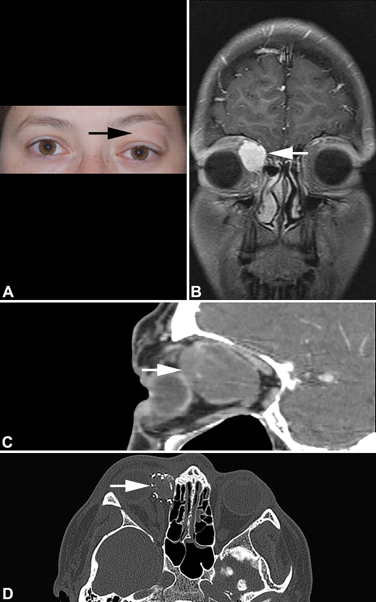
Solitary fibrous tumors (SFTs) of the orbit are rare. In order to further characterize the clinical and pathologic features of solitary fibrous tumor arising at this anatomic site, 12 cases of orbital SFTs were analyzed in conjunction with a review of 263 cases reported from the English literature in order to develop a risk prediction model. SFTs of the orbit were equally distributed between males (n = 5) and females (n = 7) with a mean patient age of 46.8 years (median 44.5 years; range 18-76 years) at initial diagnosis. The patients typically presented with swelling or mass around the orbit, with proptosis (n = 10), ptosis (n = 5), and visual changes (n = 6). Tumors were orbital (n = 10) or upper eyelid (n = 2). Mean tumor size was 2.5 cm (median 2.6 cm). Microscopically, the tumors were characterized by cytologically bland spindle cells with patternless growth, hypocellular and hypercellular areas, variable amounts of collagen, and ectatic, branching blood vessels. By immunohistochemistry, all cases had a strong nuclear STAT6 expression. All patients were initially managed with excision or biopsy, three with presurgical embolization. The two patients with biopsy only had persistent disease (mean 37.2 months), but a third patient developed distant bone metastasis at 86.9 months. Overall mean follow-up was 73.1 months: 9 patients are alive or dead without disease (mean 77.9 months), two patients with persistent disease, and one patient with metastatic disease at last follow-up (102 months). Incorporating cases sufficiently reported in the literature, a risk prediction model based on age > 45 years, tumor size > 3 cm, tumor necrosis, mitoses of > 4/2 mm2, moderate to high cellularity, and moderate to severe pleomorphism allows for risk stratification for the development of local recurrence and distant metastasis. In conclusion, orbital SFTs are rare, but can be reliably diagnosed based on the presence of characteristic morphologic features and STAT6 immunohistochemistry. Orbital tumors tend to show a higher frequency of local recurrence than distant metastasis, which can be predicted by a risk stratification model unique to orbital tumors. With late disease common, long term clinical follow-up is recommended.
Keywords: Follow-up studies; Immunohistochemistry; Orbital neoplasms; Risk assessment; STAT6; Solitary fibrous tumors.
Conflict of interest statement
All authors declare that they have no conflict of interest as it relates to this research project
Figures


References
-
- Klemperer P, Rabin CB. Primary neoplasms of the pleura: a report of five cases. Arch Pathol. 1931;11:385–412.
-
- Gengler C, Guillou L. Solitary fibrous tumour and haemangiopericytoma: evolution of a concept. Histopathology. 2006;48(1):63–74. - PubMed
-
- Mohajeri A, Tayebwa J, Collin A, et al. Comprehensive genetic analysis identifies a pathognomonic NAB2/STAT6 fusion gene, nonrandom secondary genomic imbalances, and a characteristic gene expression profile in solitary fibrous tumor. Genes Chromosomes Cancer. 2013;52(10):873–886. - PubMed
Publication types
MeSH terms
LinkOut - more resources
Full Text Sources
Research Materials
Miscellaneous
