. 2020 Dec;235(12):9785-9794.
doi: 10.1002/jcp.29792. Epub 2020 Jun 11.

Pten deletion in Dmp1-expressing cells does not rescue the osteopenic effects of Wnt/β-catenin suppression

Affiliations

Pten deletion in Dmp1-expressing cells does not rescue the osteopenic effects of Wnt/β-catenin suppression

Kyung-Eun Lim et al. J Cell Physiol. 2020 Dec.

Abstract

Skeletal homeostasis is sensitive to perturbations in Wnt signaling. Beyond its role in the bone, Wnt is a major target for pharmaceutical inhibition in a wide range of diseases, most notably cancers. Numerous clinical trials for Wnt-based candidates are currently underway, and Wnt inhibitors will likely soon be approved for clinical use. Given the bone-suppressive effects accompanying Wnt inhibition, there is a need to expose alternate pathways/molecules that can be targeted to counter the deleterious effects of Wnt inhibition on bone properties. Activation of the Pi3k/Akt pathway via Pten deletion is one possible osteoanabolic pathway to exploit. We investigated whether the osteopenic effects of β-catenin deletion from bone cells could be rescued by Pten deletion in the same cells. Mice carrying floxed alleles for Pten and β-catenin were bred to Dmp1-Cre mice to delete Pten alone, β-catenin alone, or both genes from the late-stage osteoblast/osteocyte population. The mice were assessed for bone mass, density, strength, and formation parameters to evaluate the potential rescue effect of Pten deletion in Wnt-impaired mice. Pten deletion resulted in high bone mass and β-catenin deletion resulted in low bone mass. Compound mutants had bone properties similar to β-catenin mutant mice, or surprisingly in some assays, were further compromised beyond β-catenin mutants. Pten inhibition, or one of its downstream nodes, is unlikely to protect against the bone-wasting effects of Wnt/βcat inhibition. Other avenues for preserving bone mass in the presence of Wnt inhibition should be explored to alleviate the skeletal side effects of Wnt inhibitor-based therapies.

Keywords: Akt; Pten; Wnt; osteoporosis; β-catenin.


Figures

Figure 1:
Figure 1:
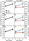
Serial in vivo DXA scans of Cre-negative (panels A-C) and 10kbDmp1-Cre positive (panels D-F) male mice carrying homozygous floxed loss-of-function Pten alleles, homozygous floxed loss-of-function β-catenin alleles, homozygous loss-of-function alleles at both loci, or WT (no floxed alleles). Scans were collected at 4 and 6 wks of age and analyzed for (A & D) whole body BMD, (B & E) BMD of the right hindlimb distal to the acetabulum, and (C & F) lumbar spine BMD. *=significantly different from WT mice at p<0.05; #=significantly different from Pten mutant mice at p<0.05; †=significantly different from βcat mutant mice at p<0.05. n=11-22/group. Female mice are depicted in Figure S2.
Figure 2:
Figure 2:
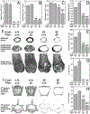
μCT-derived measurement of the distal femur metaphyseal cancellous bone, mid-femur cortical bone, and 5th lumbar vertebral cancellous bone from 10kbDmp1-Cre positive 7-wk-old male mice carrying homozygous floxed loss-of-function Pten alleles, homozygous floxed loss-of-function β-catenin alleles, homozygous floxed loss-of-function alleles at both loci, or WT (no floxed alleles). Quantitative differences in distal femur metaphyseal trabecular bone volume fraction (BV/TV; A), trabecular number (Tb.N; B) and trabecular thickness (Tb.Th; C), and in midshaft femur cortical bone volume (BV; D) and thickness (Ct.Th; E). (F) Representative 3D reconstructions of (top row) the midshaft femur, (middle row) the distal metaphysis (proximal view), and (bottom row) the caudal half of the distal femur (the ventral half was digitally removed). Quantitative differences in 5th lumbar vertebral cancellous bone volume fraction (G), and trabecular thickness (H). (I) Representative 3D reconstructions of the 5th lumbar vertebral body from all four genotypes, illustrating a 0.5 mm-thick slab through the central region of the anterio-posterior plane (top row) and the cranio-caudal plane (bottom row). *=significantly different from WT mice at p<0.05; #=significantly different from Pten mutant mice at p<0.05; †=significantly different from βcat mutant mice at p<0.05. n=6-19/group. Female mice are depicted in Figure S3.
Figure 3:
Figure 3:
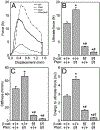
(A) Representative force–displacement curves from monotonic 3-point bending tests to failure conducted on whole femora from 7-wk-old 10kbDmp1-Cre positive male mice carrying homozygous floxed loss-of-function Pten alleles, homozygous floxed loss-of-function β-catenin alleles, homozygous floxed loss-of-function alleles at both loci, or WT (no floxed alleles). Quantification of (B) ultimate force, (C) stiffness, and (D) energy to ultimate force in all four genotypes. *=significantly different from WT mice at p<0.05; #=significantly different from Pten mutant mice at p<0.05; †=significantly different from βcat mutant mice at p<0.05. n=5-14/group.
Figure 4:
Figure 4:
Quantitative bone histomorphometry of the midshaft femur in 7-wk-old 10kbDmp1-Cre positive male mice carrying homozygous floxed loss-of-function Pten alleles, homozygous floxed loss-of-function β-catenin alleles, homozygous floxed loss-of-function alleles at both loci, or WT (no floxed alleles), measured using labels injected at 4 (green) and 6 (orange) weeks of age. Panels in (A) are magnifications of the indicated regions in (B) the whole bone images, which illustrate the extent of labeled surface on periosteal and endocortical regions. Quantification of (C) periosteal mineralizing surface per unit bone surface (MS/BS); (D) periosteal mineral apposition rate (MAR); and (E) bone formation rate per unit bone surface (BFR/BS). (F-H) Quantification of MS/BS, MAR, and BFR/BS on the endocortical surface. *=significantly different from WT mice at p<0.05; #=significantly different from Pten mutant mice at p<0.05; †=significantly different from βcat mutant mice at p<0.05. n=6-10/group.
Figure 5:
Figure 5:
ELISA-based quantification the bone resorption marker carboxy-terminal collagen crosslinks (CTx) from 6-wk-old 10kbDmp1-Cre positive male mice carrying homozygous floxed loss-of-function Pten alleles, homozygous floxed loss-of-function β-catenin alleles, homozygous floxed loss-of-function alleles at both loci, or WT (+/+; no floxed alleles). *=significantly different from WT mice at p<0.05; #=significantly different from Pten mutant mice at p<0.05. n=4–6/group.
Figure 6:
Figure 6:
(Top) Western blot of cortical bone tissue protein extract from 6-wk-old Pten floxed mice, with either WT (+/+) or floxed (f/f) βcat alleles, with (+) or without (−) Cre. The blot was immunoreacted for phosphorylated Akt, and shows activation of Akt only in the Cre-positive, βcat WT mice. (Bottom) Lane loading equivalency was assessed by Ponceau-S staining of the membrane for total protein before blotting.

References

    1. Bononi A, Bonora M, Marchi S, Missiroli S, Poletti F, Giorgi C, … Pinton P (2013). Identification of PTEN at the ER and MAMs and its regulation of Ca(2+) signaling and apoptosis in a protein phosphatase-dependent manner. Cell Death Differ, 20(12), 1631–1643. doi:10.1038/cdd.2013.77 - DOI - PMC - PubMed
    1. Brault V, Moore R, Kutsch S, Ishibashi M, Rowitch DH, McMahon AP, … Kemler R (2001). Inactivation of the beta-catenin gene by Wnt1-Cre-mediated deletion results in dramatic brain malformation and failure of craniofacial development. Development, 128(8), 1253–1264. - PubMed
    1. Burgers TA, Hoffmann MF, Collins CJ, Zahatnansky J, Alvarado MA, Morris MR, … Williams BO (2013). Mice lacking pten in osteoblasts have improved intramembranous and late endochondral fracture healing. PLoS One, 8(5), e63857. doi:10.1371/journal.pone.0063857 - DOI - PMC - PubMed
    1. Cao J, Wan L, Hacker E, Dai X, Lenna S, Jimenez-Cervantes C, … Cui R (2013). MC1R is a potent regulator of PTEN after UV exposure in melanocytes. Mol Cell, 51(4), 409–422. doi:10.1016/j.molcel.2013.08.010 - DOI - PMC - PubMed
    1. Castillo AB, Blundo JT, Chen JC, Lee KL, Yereddi NR, Jang E, … Jacobs CR (2012). Focal adhesion kinase plays a role in osteoblast mechanotransduction in vitro but does not affect load-induced bone formation in vivo. PLoS One, 7(9), e43291. doi:10.1371/journal.pone.0043291 - DOI - PMC - PubMed

Publication types

MeSH terms