Clinical Trial
. 2020 Dec;9(1):1497-1505.
doi: 10.1080/22221751.2020.1780951.

Serological differentiation between COVID-19 and SARS infections

Affiliations
Clinical Trial

Serological differentiation between COVID-19 and SARS infections

Wan Ni Chia et al. Emerg Microbes Infect. 2020 Dec.

Abstract

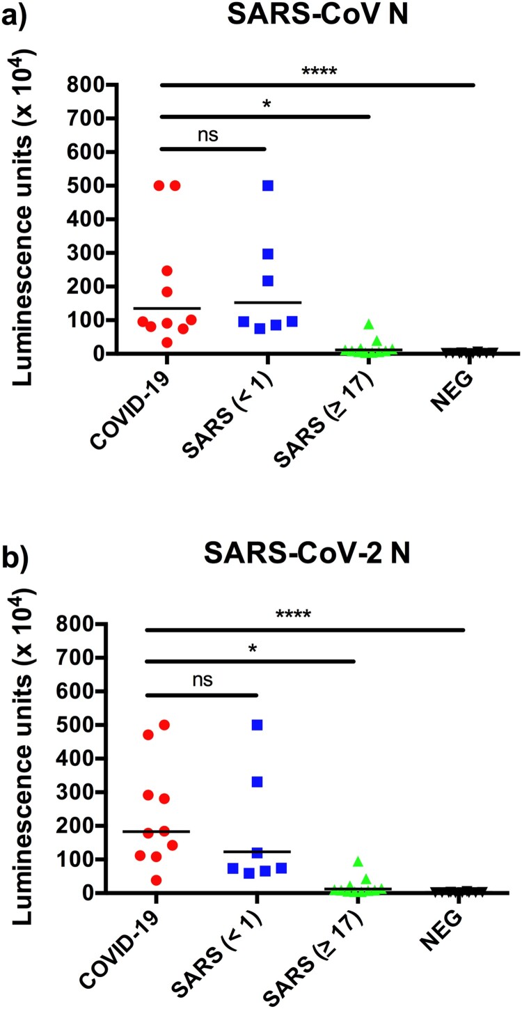
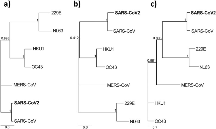
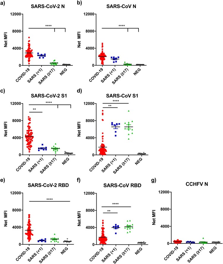
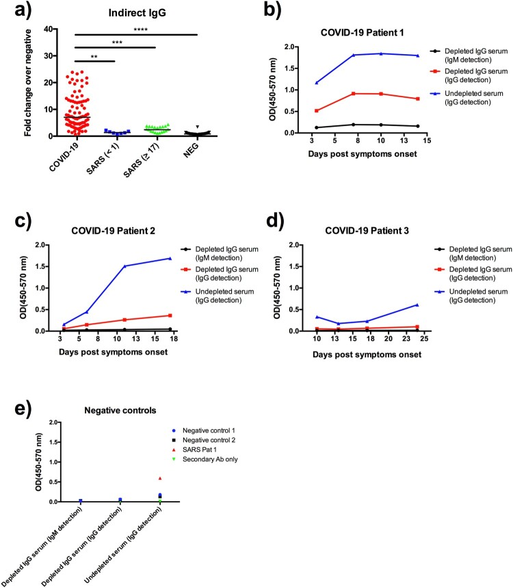
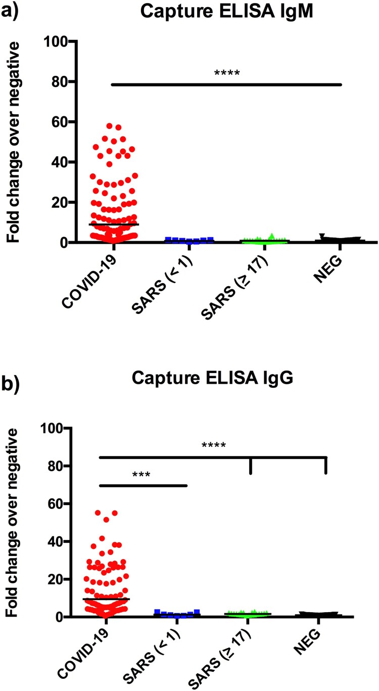
In response to the coronavirus disease 2019 (COVID-19) outbreak, caused by SARS-CoV-2, multiple diagnostic tests are required for acute disease diagnosis, contact tracing, monitoring asymptomatic infection rates and assessing herd immunity. While PCR remains the frontline test of choice in the acute diagnostic setting, serological tests are urgently needed. Unlike PCR tests which are highly specific, cross-reactivity is a major challenge for COVID-19 antibody tests considering there are six other coronaviruses known to infect humans. SARS-CoV is genetically related to SARS-CoV-2 sharing approximately 80% sequence identity and both belong to the species SARS related coronavirus in the genus Betacoronavirus of family Coronaviridae. We developed and compared the performance of four different serological tests to comprehensively assess the cross-reactivity between COVID-19 and SARS patient sera. There is significant cross-reactivity when N protein of either virus is used. The S1 or RBD regions from the spike (S) protein offers better specificity. Amongst the different platforms, capture ELISA performed best. We found that SARS survivors all have significant levels of antibodies remaining in their blood 17 years after infection. Anti-N antibodies waned more than anti-RBD antibodies, and the latter is known to play a more important role in providing protective immunity.

Keywords: COVID-19; SARS; SARS-CoV-2; antibody; serology.


Conflict of interest statement

No potential conflict of interest was reported by the author(s).

Figures

Figure 1.
Figure 1.
Rapid detection of N-specific antibodies using LIPS. Data presented are luminescence units against N proteins of SARS-CoV (a) and SARS-CoV-2 (b). The SARS sera were divided into those collected in 2003 (<1 year) or 2020 (≥17 years).
Figure 2.
Figure 2.
Phylogeny based on the three different proteins of seven known human coronaviruses. Maximum likelihood phylogenetic tree based on amino acid sequences of (a) Nucleocapsid protein (b) S1 protein and (c) RBD of human coronaviruses with 1000 bootstrap replicates. Support values are indicated at nodes. The scale bars represent substitutions per amino acid position.
Figure 3.
Figure 3.
Analysis of antibody binding from COVID-19 and SARS sera against seven recombinant proteins. Show on the Y-axis are net mean florescence (MFI) units normalised with those obtained from anti-His antibody readings for each protein (see Methods for detail). The serum panels includes 37 healthy controls, 74 sera from PCR positive COVID-19 patients, and 18 recovered SARS patients. The SARS sera were divided into those sampled in 2003 (<1 year, n=7) or 2020 (≥17 years n=11).
Figure 4.
Figure 4.
Detection of anti-RBD IgG and IgM antibodies by indirect ELISA. (a) IgG data obtained from the same serum panels as those in Figure 3. IgM testing with or without IgG depletion from three representative COVID-19 patient sera known to have high (b), medium (c) and low (d) IgG antibody levels. Also included are two healthy controls and one SARS patient serum (e). Data are presented as fold of change (Fc) over the average reading of negative controls.
Figure 5.
Figure 5.
Detection of anti-RBD IgG (a) and IgM (b) antibodies by capture ELISA. Data are presented as fold of change (Fc) over the average reading of negative controls. The same serum panels as in Figure 3 were used in this study.

References

    1. Wang C, et al. . A novel coronavirus outbreak of global health concern. Lancet. 2020;395(10223):470–473. - PMC - PubMed
    1. Coronaviridae Study Group of the International Committee on Taxonomy of, V. The species severe acute respiratory syndrome-related coronavirus: classifying 2019-nCoV and naming it SARS-CoV-2. Nat Microbiol. 2020;5(4):536–544. WHO, COVID-19 status report. https://www.who.int/emergencies/diseases/novel-coronavirus-2019/situatio...., 2020. Accessed on 1 June 2020. - PMC - PubMed
    1. Tang YW, et al. . The laboratory diagnosis of COVID-19 infection: current issues and challenges. J Clin Microbiol. 2020;58(6):e00512–20. - PMC - PubMed
    1. Petherick A. Developing antibody tests for SARS-CoV-2. Lancet. 2020;395(10230):1101–1102. - PMC - PubMed
    1. van der Hoek L, Pyrc K, Berkhout B.. Human coronavirus NL63, a new respiratory virus. FEMS Microbiol Rev. 2006;30(5):760–773. - PMC - PubMed

MeSH terms

Substances