Observational Study
. 2020 Aug;111(8):2895-2906.
doi: 10.1111/cas.14523. Epub 2020 Jul 6.

Tumor-associated macrophage-derived inflammatory cytokine enhances malignant potential of malignant pleural mesothelioma

Affiliations
Observational Study

Tumor-associated macrophage-derived inflammatory cytokine enhances malignant potential of malignant pleural mesothelioma

Daisuke Horio et al. Cancer Sci. 2020 Aug.

Abstract

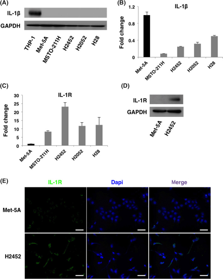
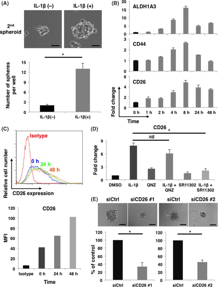
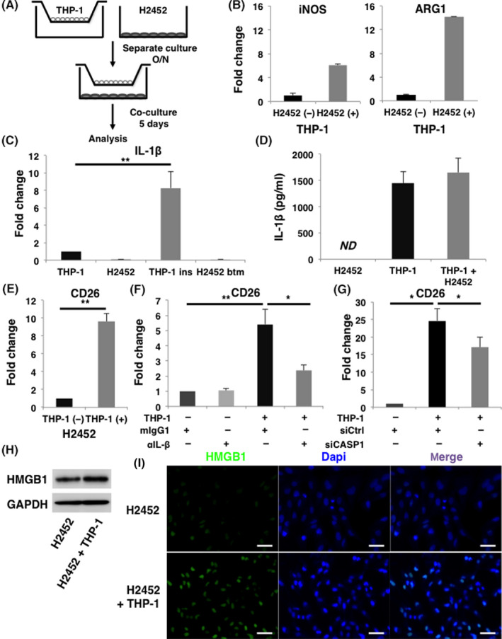
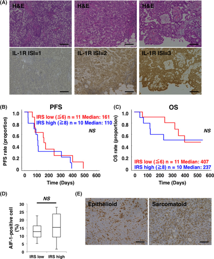
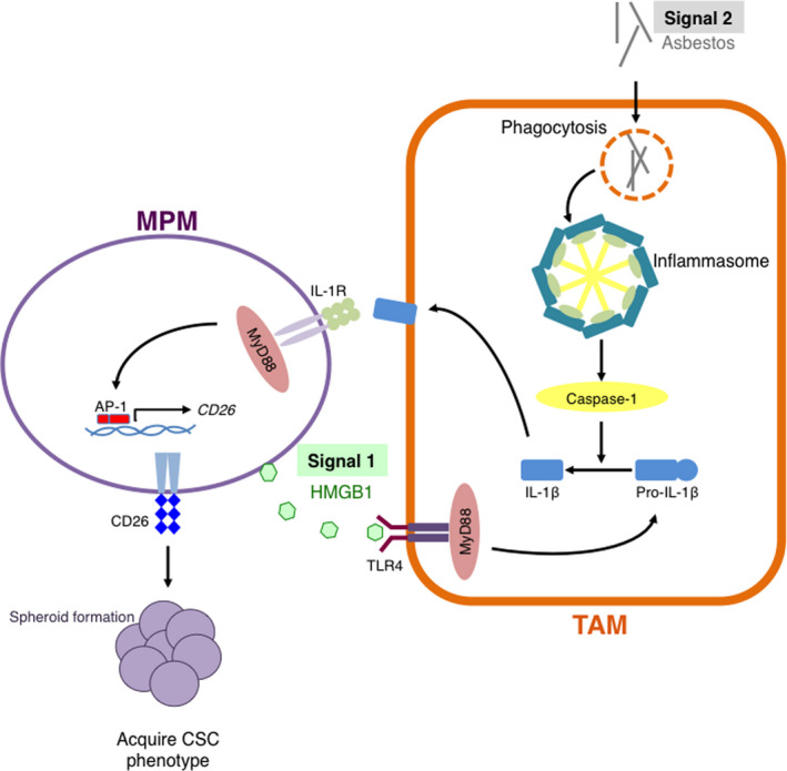
Malignant pleural mesothelioma (MPM) is an asbestos-related aggressive malignant neoplasm. Due to the difficulty of achieving curative surgical resection in most patients with MPM, a combination chemotherapy of cisplatin and pemetrexed has been the only approved regimen proven to improve the prognosis of MPM. However, the median overall survival time is at most 12 mo even with this regimen. There has been therefore a pressing need to develop a novel chemotherapeutic strategy to bring about a better outcome for MPM. We found that expression of interleukin-1 receptor (IL-1R) was upregulated in MPM cells compared with normal mesothelial cells. We also investigated the biological significance of the interaction between pro-inflammatory cytokine IL-1β and the IL-1R in MPM cells. Stimulation by IL-1β promoted MPM cells to form spheroids along with upregulating a cancer stem cell marker CD26. We also identified tumor-associated macrophages (TAMs) as the major source of IL-1β in the MPM microenvironment. Both high mobility group box 1 derived from MPM cells and the asbestos-activated inflammasome in TAMs induced the production of IL-1β, which resulted in enhancement of the malignant potential of MPM. We further performed immunohistochemical analysis using clinical MPM samples obtained from patients who were treated with the combination of platinum plus pemetrexed, and found that the overexpression of IL-1R tended to correlate with poor overall survival. In conclusion, the interaction between MPM cells and TAMs through a IL-1β/IL-1R signal could be a promising candidate as the target for novel treatment of MPM (Hyogo College of Medicine clinical trial registration number: 2973).

Keywords: IL-1β; asbestos; inflammasome; malignant pleural mesothelioma; tumor-associated macrophage.


Conflict of interest statement

No potential conflicts of interest were disclosed.

Figures

FIGURE 1
FIGURE 1
IL‐1R is overexpressed in malignant pleural mesothelioma (MPM) cells compared with mesothelial cells. A, Comprehensive analysis of IL‐1β expression in human mesothelial cell (Met‐5A) and MPM cell lines (MSTO‐211H, H2452, H2052, and H28) by immunoblotting. Cell lysates from THP‐1 cells were loaded as a positive control. Expression of IL‐1β was detected neither in mesothelial cell nor in MPM cells. B, Comprehensive analysis of IL‐1β expression in human mesothelial cell and MPM cell lines by qRT‐PCR. Expression of IL‐1β transcripts was less in all the MPM cells than in mesothelial cells. C, Comprehensive analysis of IL‐1R expression in human mesothelial cells and MPM cells lines by qRT‐PCR. Expression of IL‐1R was higher in MPM cells than in mesothelial cells. D, E, Protein level of IL‐1R in human mesothelial cell (Met‐5A) and MPM cell (H2452) was examined by immunoblotting and immunofluorescence. Consistent with the results of qRT‐PCR and immunoblotting, expression of IL‐1R was higher in H2452 cells. All the above experiments were performed at least twice with similar results. Representative data are shown. Scale Bars, 100 μm
FIGURE 2
FIGURE 2
IL‐1β promotes sphere formation through upregulation of CD26 in IL‐1R‐overexpressing H2452 cells. A, H2452 cells were cultured with or without 10 ng/mL of rhIL‐1β on poly‐HEMA plate to evaluate sphere forming ability. Representative phase contrast images and quantification of the number of spheres are shown in upper and lower panel, respectively. Results represent the mean ± SEM of 3 independent experiments performed in triplicates. Scale bars, 50 μm, *P < .01. B, Representative data of expression of ALDH1A3, CD44, and CD26 transcripts induced by rhIL‐1β at the indicated time points are shown. Experiments were repeated twice in triplicates with similar results. Error bars, SEM. C, Cell surface CD26 expression induced by 10 ng/mL of rhIL‐1β at the indicated time points was analyzed by flow cytometry. Representative histogram and mean fluorescence intensity (MFI) are shown in upper and lower panel, respectively. Experiments were repeated twice with similar results. Error bars, SEM. D, Expression of CD26 transcripts in H2452 cells stimulated by 10 ng/mL of rhIL‐1β for 8 h in the presence of QNZ or SR11302 was measured by qRT‐PCR. Results represent mean ± SEM of 3 independent experiments performed in triplicates. *P < .01. E, Sphere forming ability of H2452 cells either transfected with control or 2 different CD26 targeting siRNAs (siCD26 #1 and siCD26 #2) was examined. Representative phase contrast images and quantification of the relative number of spheres are shown in left and right panel, respectively. Results represent mean ± SEM of 3 independent experiments performed in triplicates. Scale bars, 100 μm, *P < .01
FIGURE 3
FIGURE 3
IL‐1β derived from macrophages induces CD26 expression in malignant pleural mesothelioma (MPM) cells. A, Schema of coincubation assay. THP‐1‐derived macrophages and H2452 cells were coincubated for 5 d and separately analyzed. B, Expression of transcripts of M1 marker iNOS and M2 marker ARG1 in THP‐1‐derived macrophages in the absence or presence of coincubating with H2452 cells are shown in the left and right panels, respectively. C, Amounts of IL‐1β transcripts in H2452 cells and THP‐1‐derived macrophages were measured by qRT‐PCR. Results of the samples of THP‐1‐derived macrophages in the insert (THP‐1 ins) and H2452 cells in the bottom well (H2452 btm) harvested after coincubation using Transwell (Costar) inserts were presented in the third and fourth lane from the left, respectively. D, ELISA analysis about the concentration of IL‐1β in the culture medium. Samples were collected from the supernatants of H2452 cells, THP‐1‐derived macrophages, and co‐culture medium of these cells. ND, not detected. E, Amount of CD26 transcripts in H2452 cells under coincubation with THP‐1‐derived macrophages was also analyzed by qRT‐PCR. F, Expression of CD26 transcripts in H2452 cells when coincubated with THP‐1‐derived macrophage in the presence of 1 μg/mL of anti‐IL‐1β neutralizing Ab (αIL‐1β) or its isotype control mouse IgG1 (mIgG1) was evaluated. G, CD26 gene expression in H2452 cells was examined when coincubated with either control or CASP1 siRNA‐transfected THP‐1‐derived macrophages. H, Expression of HMGB1 in H2452 cells under coincubation with THP‐1‐derived macrophages was examined by immunoblotting. I, Representative immunofluorescence images are shown of HMGB1 expression in H2452 cells when they were coincubated with THP‐1‐derived macrophages. All the experiments were performed at least 3 times. Quantification data represent mean ± SEM. *P < .05, **P < .01, Scale bars, 50 μm
FIGURE 4
FIGURE 4
Overexpression of IL‐1R may be a negative prognostic factor in patients with malignant pleural mesothelioma (MPM) who were treated with platinum plus pemetrexed. A, Representative images of hematoxylin‐eosin staining and IL‐1R IHC staining of clinical samples from patients with MPM are shown in the upper panels and lower panels, respectively. Immunostaining intensity (ISI) for IL‐1R was scored from negative to strong. Mild staining (ISI = 1), moderate staining (ISI = 2), and strong staining (ISI = 3) were indicated in lower left, center, and right panel, respectively. Scale bars, 100 μm. B, Kaplan‐Meier curve for progression‐free survival (PFS) between the immunoreactive score (IRS) low group (n = 11, red) and the high group (n = 10, blue). C, Kaplan‐Meier curve for OS between IRS score low group (n = 11, red) and high group (n = 10, blue). NS, not significant. D, Comparison of AIF‐1‐positive cell ratio in MPM tissue between the IRS low group (n = 11) and IRS high group (n = 10). The middle line, the upper end, and the lower end of the box plot represent median, 75% and 25% values, respectively. NS, not significant. E, Representative images of AIF‐1 immunohistochemistry (IHC) staining of epithelioid and sarcomatoid MPM samples were shown in the left panel and right panel, respectively. Scale bars, 100 μm
FIGURE 5
FIGURE 5
Tumor‐associated macrophages (TAMs) contribute in the acquisition of a CSC‐like phenotype in malignant pleural mesothelioma (MPM) cells via IL‐1β/IL‐1R axis. HMGB1 released from MPM cells (signal 1) induces pro‐IL‐1β production through activating TLR4 in TAMs. In TAMs, phagocytosed asbestos constitutively activates the inflammasome (signal 2), which in turn causes maturation and secretion of IL‐1β. Secreted IL‐1β interacts with IL‐1R on MPM cells via a paracrine mechanism. Finally, MPM cells acquire a cancer stem cell (CSC)‐like phenotype

References

    1. Ricciardi S, Cardillo G, Zirafa CC, et al. Surgery for malignant pleural mesothelioma: an international guidelines review. J Thorac Dis. 2018;10(Suppl 2):S285‐S292. - PMC - PubMed
    1. Scherpereel A, Wallyn F, Albelda SM, Munck C. Novel therapies for malignant pleural mesothelioma. Lancet Oncol. 2018;19(3):e161‐e172. - PubMed
    1. Bibby AC, Tsim S, Kanellakis N, et al. Malignant pleural mesothelioma: an update on investigation, diagnosis and treatment. Eur Respir Rev. 2016;25(142):472‐486. - PMC - PubMed
    1. Quispel‐Janssen J, van der Noort V, de Vries JF, et al. Programmed death 1 blockade with nivolumab in patients with recurrent malignant pleural mesothelioma. J Thorac Oncol. 2018;13(10):1569‐1576. - PubMed
    1. Scherpereel A, Mazieres J, Greillier L, et al. Nivolumab or nivolumab plus ipilimumab in patients with relapsed malignant pleural mesothelioma (IFCT‐1501 MAPS2): a multicentre, open‐label, randomised, non‐comparative, phase 2 trial. Lancet Oncol. 2019;20(2):239‐253. - PubMed

Publication types

MeSH terms