. 2020 Jun 10;21(11):4137.
doi: 10.3390/ijms21114137.

Cerebellar Blood Flow and Gene Expression in Crossed Cerebellar Diaschisis after Transient Middle Cerebral Artery Occlusion in Rats

Affiliations

Cerebellar Blood Flow and Gene Expression in Crossed Cerebellar Diaschisis after Transient Middle Cerebral Artery Occlusion in Rats

Naoya Kidani et al. Int J Mol Sci. .

Abstract

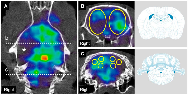
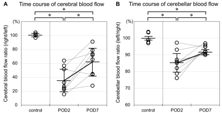
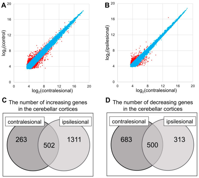
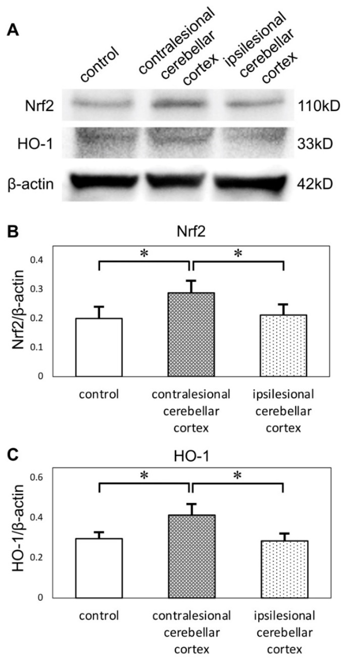
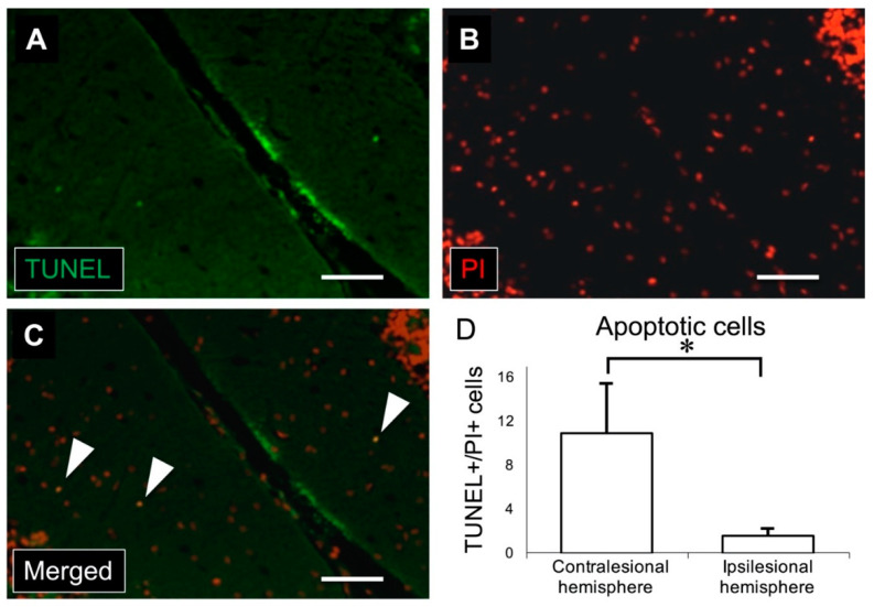
Crossed cerebellar diaschisis (CCD) is a state of hypoperfusion and hypometabolism in the contralesional cerebellar hemisphere caused by a supratentorial lesion, but its pathophysiology is not fully understood. We evaluated chronological changes in cerebellar blood flow (CbBF) and gene expressions in the cerebellum using a rat model of transient middle cerebral artery occlusion (MCAO). CbBF was analyzed at two and seven days after MCAO using single photon emission computed tomography (SPECT). DNA microarray analysis and western blotting of the cerebellar cortex were performed and apoptotic cells in the cerebellar cortex were stained. CbBF in the contralesional hemisphere was significantly decreased and this lateral imbalance recovered over one week. Gene set enrichment analysis revealed that a gene set for "oxidative phosphorylation" was significantly upregulated while fourteen other gene sets including "apoptosis", "hypoxia" and "reactive oxygen species" showed a tendency toward upregulation in the contralesional cerebellum. MCAO upregulated the expressions of nuclear factor erythroid 2-related factor 2 (Nrf2) and heme oxygenase-1 (HO-1) in the contralesional cerebellar cortex. The number of apoptotic cells increased in the molecular layer of the contralesional cerebellum. Focal cerebral ischemia in our rat MCAO model caused CCD along with enhanced expression of genes related to oxidative stress and apoptosis.

Keywords: apoptosis; cerebral blood flow; crossed cerebellar diaschisis; ischemic stroke; oxidative stress.


Conflict of interest statement

The authors declare no conflict of interest. The funders had no role in the design of the study; in the collection, analyses, or interpretation of data; in the writing of the manuscript, or in the decision to publish the results.

Figures

Figure 1
Figure 1
Single photon emission computed tomography (SPECT)/computed tomography (CT) images and regions of interest (ROI) settings to evaluate cerebral and cerebellar blood flow after MCAO. SPECT acquisitions were color-coded with National Institute of Health spectrum color scale. (A), Vertical section of SPECT/CT image obtained 2 days after middle cerebral artery occlusion (MCAO) shows obvious hypointensity at the right middle cerebral artery territory (*). Dashed line “b” indicates the coronal slice 2 mm posterior from bregma and dashed line “c” indicates the coronal slice 11 mm posterior from bregma. (B) In the coronal slice 2 mm posterior from bregma, 6 × 9 × 6 mm 3D-elliptical ROIs were symmetrically placed. (C) In the coronal slice 11 mm posterior from bregma, 2 × 2 × 2 mm global ROIs were symmetrically placed at medial cerebellar cortex, lateral cerebellar cortex and cerebellar nuclei of each side respectively.
Figure 2
Figure 2
(A) Rats in the control group showed no apparent laterality of cerebral blood flow. Cerebral blood flow ratio (right/left) was significantly decreased two and seven days after MCAO. The plots of each individual are connected with bottled lines. This ratio significantly recuperated over time. (B) Rats in the control group showed no apparent laterality of cerebellar blood flow. Cerebellar blood flow ratio (left/right) was significantly decreased two and seven days after MCAO, but this decrease was of a lesser degree than that in the cerebrum. As in the cerebrum, the decrease in blood flow ratio in the cerebellum likewise recuperated over time. (* p < 0.05, n = 5 in the control group, n = 8 in the MCAO group).
Figure 3
Figure 3
(A,B) Correlation plot of the contralesional, ipsilesional and control group. The genes with expression change between two conditions <2.0× are labeled as blue and ≥2.0× as red. (C,D) The mRNA gene expression ratio was expressed as the ratio of the fluorescence intensity of the contra-or ipsilesional group to that of the control group. Increasing genes were defined as those for which Log2 (ratio) > 1 if the ratio was more than double, while decreasing genes were defined as those for which Log2 (ratio) < −1 if the ratio was less than one-half.
Figure 4
Figure 4
(A) Western blots of cerebellar cortices show expression of Nrf2 and HO-1. Protein levels were normalized to beta-actin. (B) Quantification of western blots by densitometric analysis indicated that the expression of Nrf2 was upregulated in contralesional cerebellar cortices. (C) The expression of HO-1 was also upregulated in contralesional cerebellar cortices (* p < 0.05 versus other groups, n = 6 in each group). Nrf2: nuclear factor erythroid 2-related factor 2, HO-1: heme oxygenase-1.
Figure 5
Figure 5
(A) Immunostaining for TUNEL (green) shows apoptotic cells in the molecular layer of the cerebellar cortex. (B) Nuclei were stained with PI in red. (C) Merged image of TUNEL and PI immunostaining. White arrowheads indicate TUNEL/PI-positive cells. (D) There was a significant increase in the number of TUNEL/PI-positive cells in the contralesional (left) cerebellar cortex compared to the ipsilesional (right) cerebellar cortex (scale bar: 50 μm, * p < 0.001, n = 11 in each group). TUNEL: terminal deoxynucleotidyl transferase deoxyuridine triphosphate nick-end labeling, PI: propidium iodide.

References

    1. Finger S., Koehler P.J., Jagella C. The Monakow concept of diaschisis: Origins and perspectives. Arch. Neurol. 2004;61:283–288. doi: 10.1001/archneur.61.2.283. - DOI - PubMed
    1. Baron J.C., Bousser M.G., Comar D., Castaigne P. “Crossed cerebellar diaschisis” in human supratentorial brain infarction. Trans. Am. Neurol. Assoc. 1981;105:459–461. - PubMed
    1. Carrera E., Tononi G. Diaschisis: Past, present, future. Brain. 2014;137:2408–2422. doi: 10.1093/brain/awu101. - DOI - PubMed
    1. Gold L., Lauritzen M. Neuronal deactivation explains decreased cerebellar blood flow in response to focal cerebral ischemia or suppressed neocortical function. Proc. Natl. Acad. Sci. USA. 2002;99:7699–7704. doi: 10.1073/pnas.112012499. - DOI - PMC - PubMed
    1. Lim J.S., Ryu Y.H., Kim B.M., Lee J.D. Crossed cerebellar diaschisis due to intracranial hematoma in basal ganglia or thalamus. J. Nucl. Med. 1998;39:2044–2047. - PubMed

MeSH terms