. 2020 Jun 10;12(6):1742.
doi: 10.3390/nu12061742.

Attenuation of Lipopolysaccharide-Induced Acute Lung Injury by Hispolon in Mice, Through Regulating the TLR4/PI3K/Akt/mTOR and Keap1/Nrf2/HO-1 Pathways, and Suppressing Oxidative Stress-Mediated ER Stress-Induced Apoptosis and Autophagy

Affiliations

Attenuation of Lipopolysaccharide-Induced Acute Lung Injury by Hispolon in Mice, Through Regulating the TLR4/PI3K/Akt/mTOR and Keap1/Nrf2/HO-1 Pathways, and Suppressing Oxidative Stress-Mediated ER Stress-Induced Apoptosis and Autophagy

Ching-Ying Huang et al. Nutrients. .

Abstract

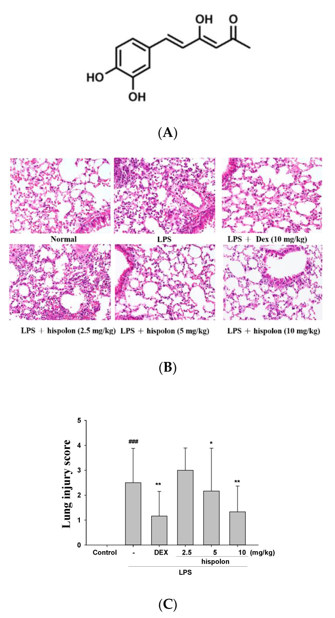
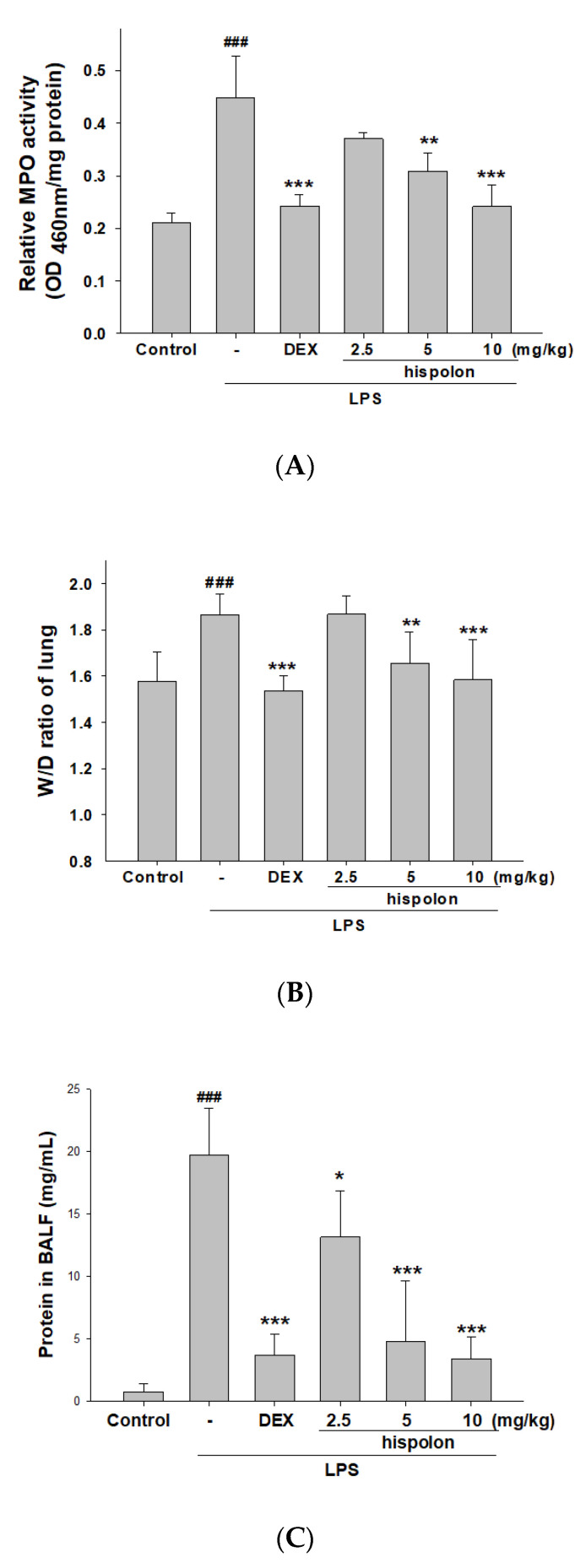
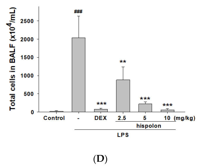
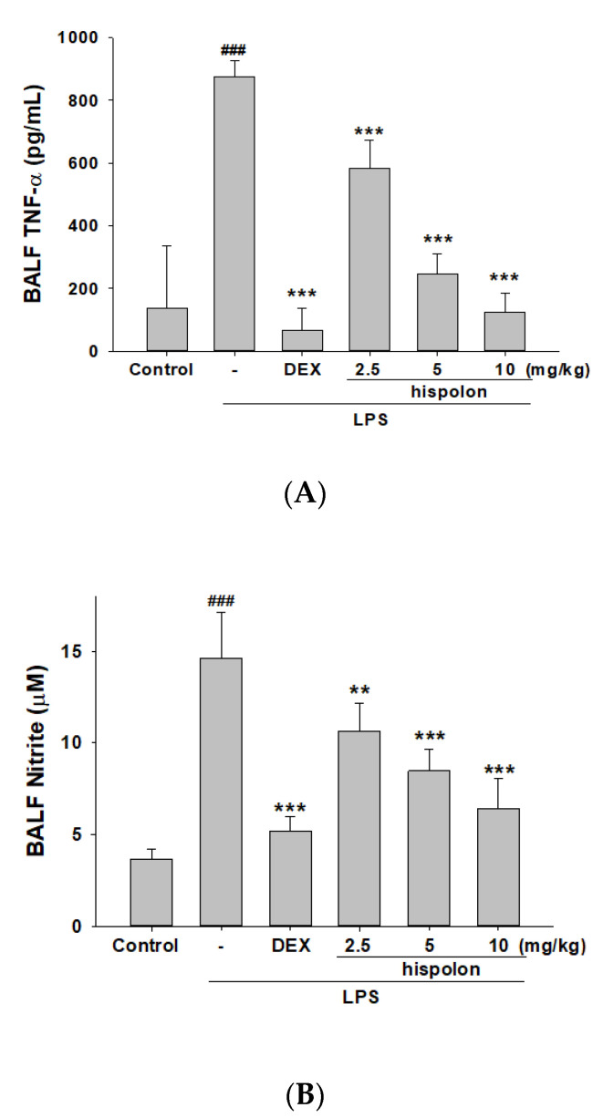
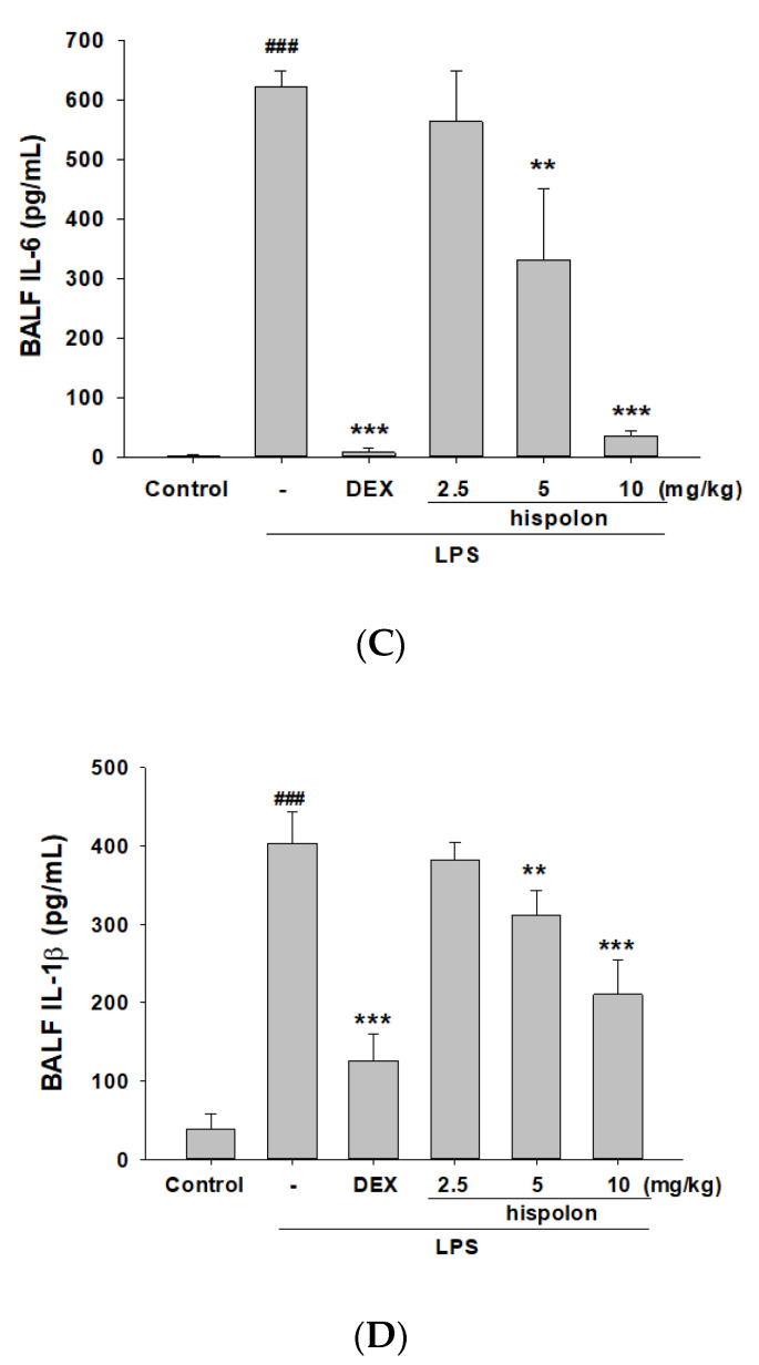
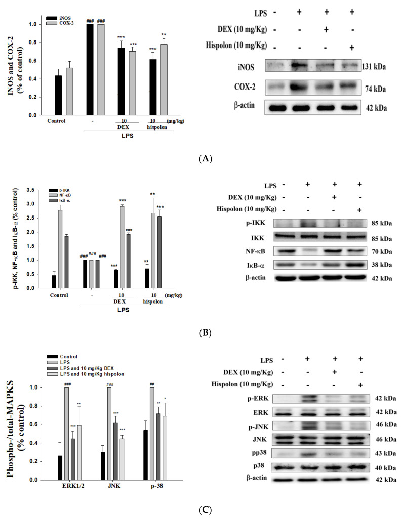
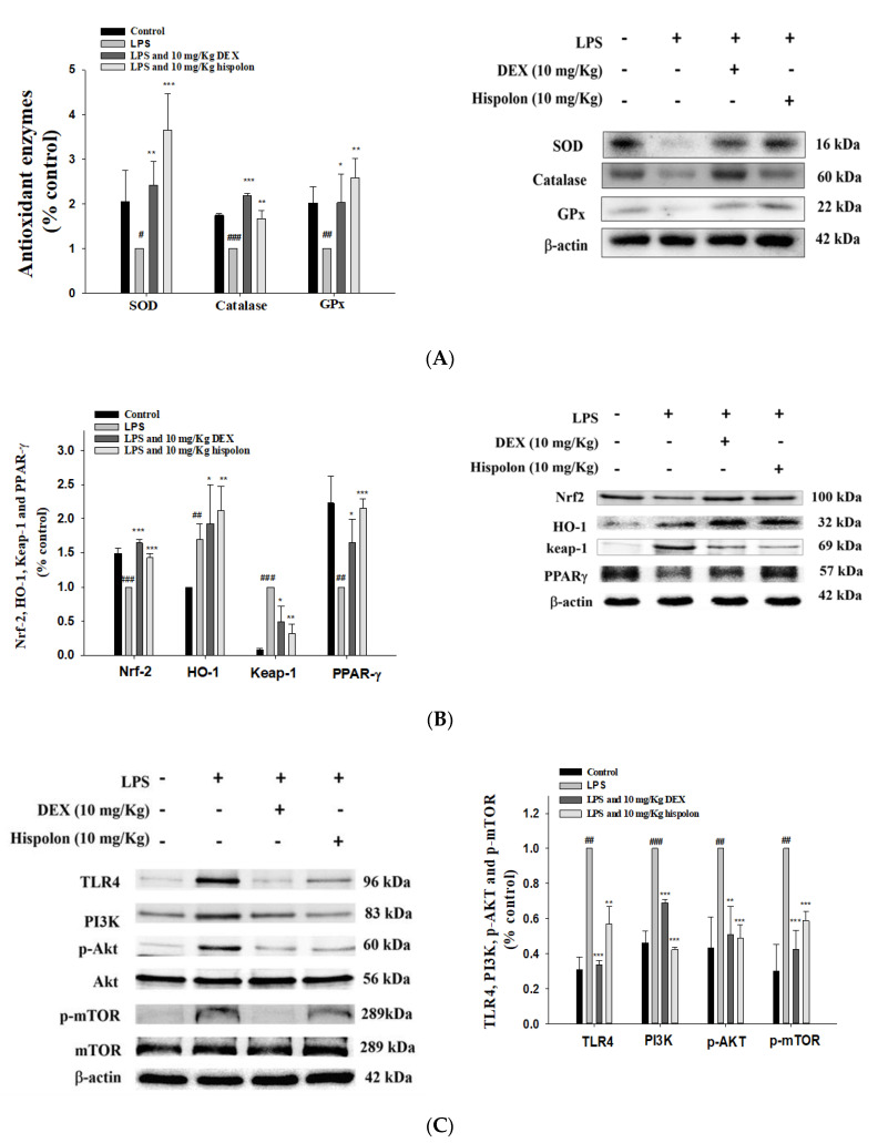
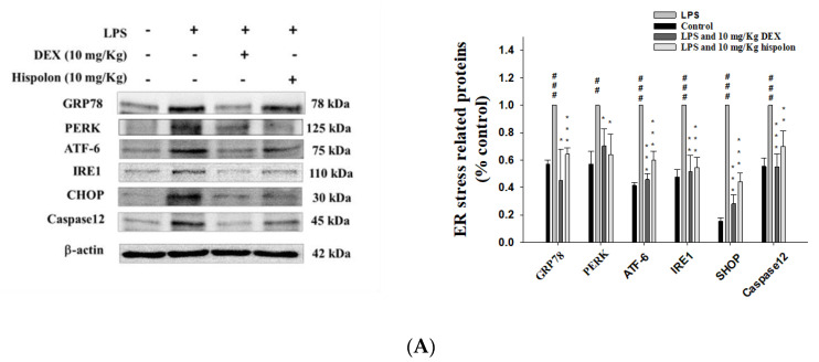
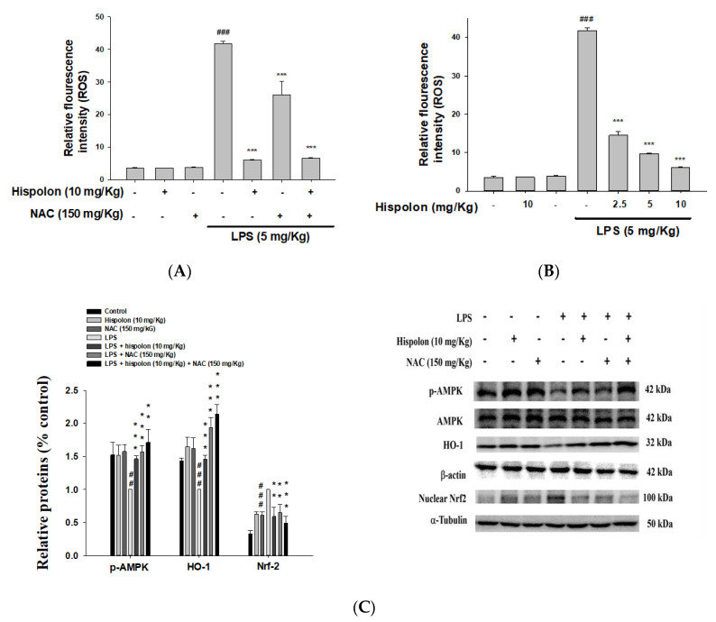
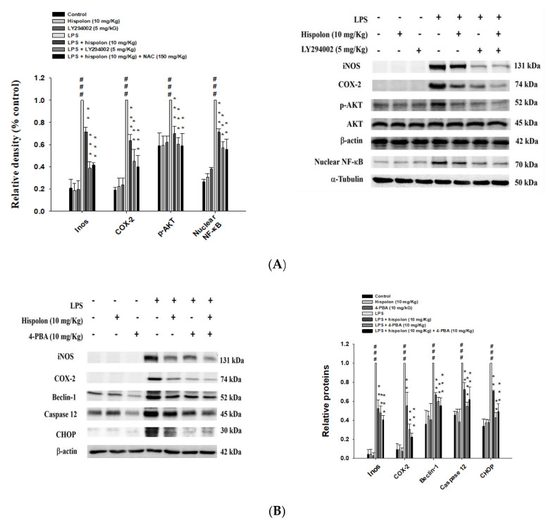
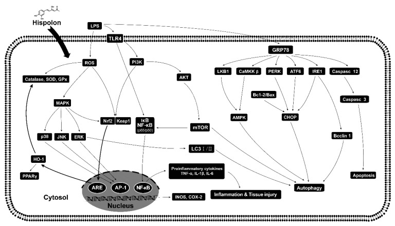
The anti-inflammatory effect of hispolon has identified it as one of the most important compounds from Sanghuangporus sanghuang. The research objectives were to study this compound using an animal model by lipopolysaccharide (LPS)-induced acute lung injury. Hispolon treatment reduced the production of the pro-inflammatory mediator NO, TNF-α, IL-1β, and IL-6 induced by LPS challenge in the lung tissues, as well as decreasing their histological alterations and protein content. Total cell number was also reduced in the bronchoalveolar lavage fluid (BALF). Moreover, hispolon inhibited iNOS, COX-2 and IκB-α and phosphorylated IKK and MAPK, while increasing catalase, SOD, GPx, TLR4, AKT, HO-1, Nrf-2, Keap1 and PPARγ expression, after LPS challenge. It also regulated apoptosis, ER stress and the autophagy signal transduction pathway. The results of this study show that hispolon regulates LPS-induced ER stress (increasing CHOP, PERK, IRE1, ATF6 and GRP78 protein expression), apoptosis (decreasing caspase-3 and Bax and increasing Bcl-2 expression) and autophagy (reducing LC3 I/II and Beclin-1 expression). This in vivo experimental study suggests that hispolon suppresses the LPS-induced activation of inflammatory pathways, oxidative injury, ER stress, apoptosis and autophagy and has the potential to be used therapeutically in major anterior segment lung diseases.

Keywords: ER stress; HO-1; LPS; Nrf-2; anti-inflammation; apoptosis; hispolon.


Conflict of interest statement

All authors have no conflicts of interests exists.

Figures

Figure 1
Figure 1
(A) The chemical structural formula of hispolon, (B) the lung injury scores and (C) the effects of hispolon (2.5, 5 and 10 mg/mL) on LPS-induced histopathologic alterations in lung tissues of mice. After LPS challenge, the lungs were prepared for histological assessment. Sections were stained with H&E and viewed under magnification (400×). Data are presented as the means ± S.E.M (n = 6). ### p < 0.001 versus the control group. * p < 0.5 and ** p < 0.01 versus the LPS group.
Figure 2
Figure 2
Hispolon improved (A) pulmonary edema (W/D ratio) and (B) Myeloperoxidase (MPO) activity, and decreased (C) cell counts and (D) total protein in the bronchoalveolar lavage fluid (BALF). Lung tissues were measured by calculating the W/D ratios. Total cells and total proteins of BALF were assessed. Data are presented as means ± S.E.M. (n = 6). ### p < 0.001 versus the control group. * p < 0.05, ** p < 0.01 and *** p < 0.001 versus the lipopolysaccharide (LPS) group.
Figure 2
Figure 2
Hispolon improved (A) pulmonary edema (W/D ratio) and (B) Myeloperoxidase (MPO) activity, and decreased (C) cell counts and (D) total protein in the bronchoalveolar lavage fluid (BALF). Lung tissues were measured by calculating the W/D ratios. Total cells and total proteins of BALF were assessed. Data are presented as means ± S.E.M. (n = 6). ### p < 0.001 versus the control group. * p < 0.05, ** p < 0.01 and *** p < 0.001 versus the lipopolysaccharide (LPS) group.
Figure 3
Figure 3
Hispolon decreased (A) NO, (B) TNF-α, (C) IL-1β and (D) IL-6 in the BALF. Pro-Inflammatory cytokine were measured after LPS challenge by ELISA. Data are represented as means ± S.E.M. (n = 6). ### p < 0.001 versus the control group. ** p < 0.01 and *** p < 0.001 versus the LPS group.
Figure 3
Figure 3
Hispolon decreased (A) NO, (B) TNF-α, (C) IL-1β and (D) IL-6 in the BALF. Pro-Inflammatory cytokine were measured after LPS challenge by ELISA. Data are represented as means ± S.E.M. (n = 6). ### p < 0.001 versus the control group. ** p < 0.01 and *** p < 0.001 versus the LPS group.
Figure 4
Figure 4
Effects of hispolon on the activation of (A) iNOS, COX-2, (B) IκB-α, NF-κB and (C) the phosphorylation of MAPK axis in lung tissue of LPS-induced acute lung injury (ALI) mice. Lung tissue extracts were subjected to Western blot analysis using antibodies for iNOS, COX-2, IκB-α and NF-κB and MAPK phosphorylation. Data are represented as means ± S.E.M. (n = 3). ### p < 0.001 versus the control group. * p < 0.05, ** p < 0.01 and *** p < 0.001 versus the LPS group.
Figure 5
Figure 5
Effects of hispolon on (A) LPS challenge catalase, superoxide dismutase (SOD) and GPx; (B) HO-1, Nrf2, Keap1 and PPARγ; (C) and TLR4, PI3K, Akt and mTOR protein expression in the lungs. Lung tissue extracts were subjected to Western blot analysis using antibodies for catalase, SOD, GPx, HO-1, Nrf2, Keap1, PPARγ, TLR4, PI3K, Akt and mTOR. Data are represented as means ± S.E.M. (n = 3). ## p < 0.01 and ### p < 0.001 versus the control group. * p < 0.05, ** p < 0.01 and *** p < 0.001 versus the LPS group.
Figure 6
Figure 6
Effects of hispolon on (A) ER stress relative enzymes (PERK, IRE1 ATF6, GRP94, CHOP and caspase-12), (B) autophagy related enzymes (LC3-II/I and Beclin 1), (C) LKB1/CaMKK–AMPK signaling, and (D) apoptosis-related protein (Bcl-2, Bax and caspase-3) levels in the lungs of LPS challenge ALI. Lung tissue extracts were subjected to Western blot analysis using antibodies for PERK, IRE1 ATF6, GRP94, CHOP, caspase-12, LC3-II/I, Beclin 1, LKB1, CaMKK, AMPK, Bcl-2, Bax and caspase-3. Data are represented as means ± S.E.M. (n = 3). ## p < 0.01 and ### p < 0.001 versus the control group. * p < 0.05, ** p < 0.01 and *** p < 0.001 versus the LPS group.
Figure 6
Figure 6
Effects of hispolon on (A) ER stress relative enzymes (PERK, IRE1 ATF6, GRP94, CHOP and caspase-12), (B) autophagy related enzymes (LC3-II/I and Beclin 1), (C) LKB1/CaMKK–AMPK signaling, and (D) apoptosis-related protein (Bcl-2, Bax and caspase-3) levels in the lungs of LPS challenge ALI. Lung tissue extracts were subjected to Western blot analysis using antibodies for PERK, IRE1 ATF6, GRP94, CHOP, caspase-12, LC3-II/I, Beclin 1, LKB1, CaMKK, AMPK, Bcl-2, Bax and caspase-3. Data are represented as means ± S.E.M. (n = 3). ## p < 0.01 and ### p < 0.001 versus the control group. * p < 0.05, ** p < 0.01 and *** p < 0.001 versus the LPS group.
Figure 7
Figure 7
Hispolon downregulated ROS (A), a ROS inhibitor (NAC) reduced ROS (B) in the BALF and regulated p-AMPK, HO-1 and nuclear Nrf2 (C) protein expression in the lungs. ROS was detected after LPS challenge by ELISA. Lung tissue extracts were subjected to Western blot analysis using antibodies for p-AMPK, HO-1 and nuclear Nrf2. Data are represented as means ± S.E.M. (n = 3). ## p < 0.01 and ### p < 0.001 versus the control group. ** p < 0.01 and *** p < 0.001 versus the LPS group.
Figure 8
Figure 8
Hispolon, an AKT inhibitor (LY294002) (A) and an ER inhibitor (4-PBA) (B) regulated protein expression in LPS-challenged mice. Lung tissue extracts were subjected to Western blot analysis, using antibodies for iNOS, COX-2, Beclin-1, caspase 12 and CHOP. Data are represented as means ± S.E.M. (n = 3). ### p < 0.001, compared with the control group. ** p < 0.01 and *** p < 0.001 versus the LPS group.
Figure 9
Figure 9
A scheme for the protective effect of hispolon against LPS challenge inflammation.

References

    1. Park J., Chen Y., Zheng M., Ryu J., Cho G.J., Surh Y.J., Sato D., Hamada H., Ryter S.W., Kim U.H., et al. Pterostilbene 4′-β-glucoside attenuates LPS-induced acute lung injury via induction of heme oxygenase-1. Oxid. Med. Cell. Longev. 2018;2018:2747018. doi: 10.1155/2018/2747018. - DOI - PMC - PubMed
    1. Ware L.B., Matthay M.A. The acute respiratory distress syndrome. N. Eng. J. Med. 2000;342:1334–1349. doi: 10.1056/NEJM200005043421806. - DOI - PubMed
    1. Takeda K., Akira S. Toll-like receptors in innate immunity. Int. Immunol. 2005;17:1–14. doi: 10.1093/intimm/dxh186. - DOI - PubMed
    1. Lin W.C., Deng J.S., Huang S.S., Wu S.H., Chen C.C., Lin W.R., Lin H.Y., Huang G.J. Anti-Inflammatory Activity of Sanghuangporus Sanghuang Mycelium. Int. J. Mol. Sci. 2017;18:347. doi: 10.3390/ijms18020347. - DOI - PMC - PubMed
    1. Keppler-Noreuil K.M., Parker V.E.R., Darling T.N., Martinez-Agosto J.A. Somatic overgrowth disorders of the PI3K/AKT/mTOR pathway & therapeutic strategies. Am. J. Med. Genet. C Semin. Med. Genet. 2016;172:402–421. - PMC - PubMed

MeSH terms