. 2020 Jun 10;12(6):629.
doi: 10.3390/v12060629.

The Anticoagulant Nafamostat Potently Inhibits SARS-CoV-2 S Protein-Mediated Fusion in a Cell Fusion Assay System and Viral Infection In Vitro in a Cell-Type-Dependent Manner

Affiliations

The Anticoagulant Nafamostat Potently Inhibits SARS-CoV-2 S Protein-Mediated Fusion in a Cell Fusion Assay System and Viral Infection In Vitro in a Cell-Type-Dependent Manner

Mizuki Yamamoto et al. Viruses. .

Abstract

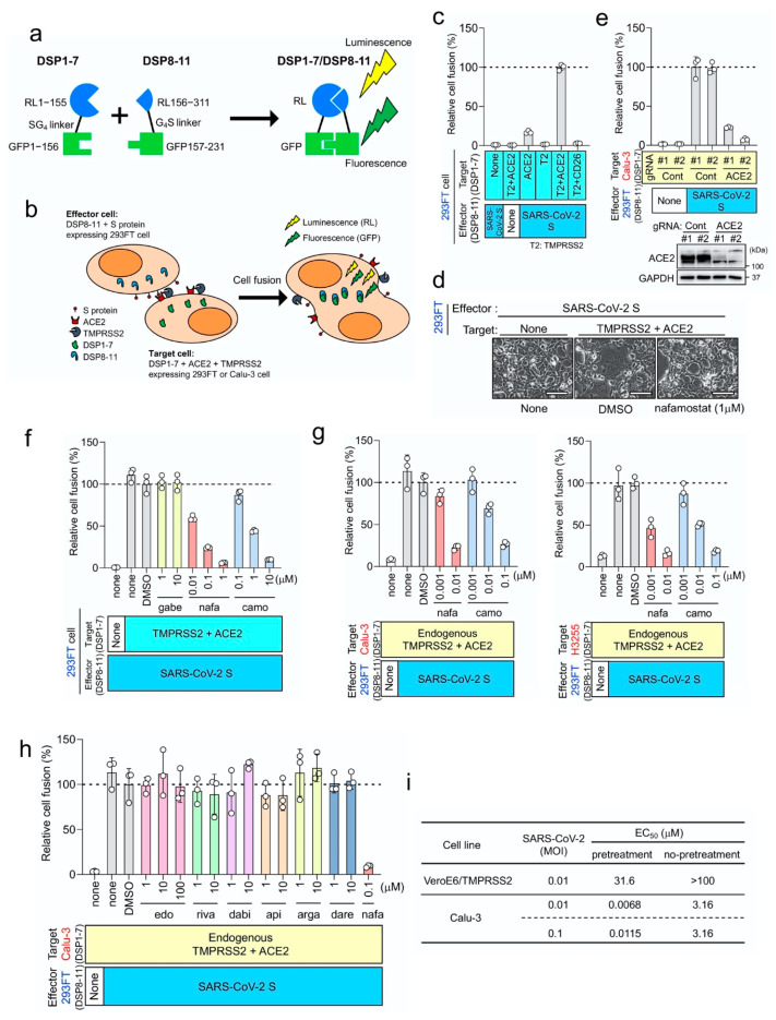
Although infection by SARS-CoV-2, the causative agent of coronavirus pneumonia disease (COVID-19), is spreading rapidly worldwide, no drug has been shown to be sufficiently effective for treating COVID-19. We previously found that nafamostat mesylate, an existing drug used for disseminated intravascular coagulation (DIC), effectively blocked Middle East respiratory syndrome coronavirus (MERS-CoV) S protein-mediated cell fusion by targeting transmembrane serine protease 2 (TMPRSS2), and inhibited MERS-CoV infection of human lung epithelium-derived Calu-3 cells. Here we established a quantitative fusion assay dependent on severe acute respiratory syndrome coronavirus 2 (SARS-CoV-2) S protein, angiotensin I converting enzyme 2 (ACE2) and TMPRSS2, and found that nafamostat mesylate potently inhibited the fusion while camostat mesylate was about 10-fold less active. Furthermore, nafamostat mesylate blocked SARS-CoV-2 infection of Calu-3 cells with an effective concentration (EC)50 around 10 nM, which is below its average blood concentration after intravenous administration through continuous infusion. On the other hand, a significantly higher dose (EC50 around 30 mM) was required for VeroE6/TMPRSS2 cells, where the TMPRSS2-independent but cathepsin-dependent endosomal infection pathway likely predominates. Together, our study shows that nafamostat mesylate potently inhibits SARS-CoV-2 S protein-mediated fusion in a cell fusion assay system and also inhibits SARS-CoV-2 infection in vitro in a cell-type-dependent manner. These findings, together with accumulated clinical data regarding nafamostat's safety, make it a likely candidate drug to treat COVID-19.

Keywords: SARS-CoV-2; TMPRSS2; fusion inhibitor.


Conflict of interest statement

The authors declare no conflict of interest.

Figures

Figure 1
Figure 1
Nafamostat mesylate potently inhibits transmembrane serine protease 2 (TMPRSS2)-dependent severe acute respiratory syndrome coronavirus 2 (SARS-CoV-2) entry into lung epithelium-derived Calu-3 cells. (a) Dual split protein (DSP)1-7 has the structure Renilla luciferase (RL)1–155-Ser-Gly-Gly-Gly-Gly-green fluorescent protein (GFP)1–156. DSP8-11 has the structure Met-RL156–311-Gly-Gly-Gly-Gly-Ser-GFP157–231. DSP1-7 and DSP8-11 reassociate efficiently, resulting in reconstitution of functional RL and GFP to generate luminescent and fluorescent signals, respectively. (b) Effector cells (293FT cells expressing DSP8-11 and S protein) and target cells (293FT or Calu-3 cells expressing DSP1-7, angiotensin I converting enzyme 2 (ACE2) and TMPRSS2) were co-cultured. Both GFP (fluorescence) and RL (luminescence) signals were generated following DSP1-7 and DSP8-11 reassociation upon cell fusion. (c) Different combinations of the effector and target cells were cocultured, and the resulting RL activity was measured. Relative cell-fusion values were calculated by normalizing the RL activity of each co-culture to that of the co-culture of cells expressing S protein with those expressing both receptor and TMPRSS2, which was set to 100%. (d) Phase contrast images of SARS-CoV-2 S protein-mediated-cell fusion. Scale bars, 100 μm. (e) The fusion assay using wild type and ACE2-kockout Calu-3 cells. (f) Three clinically used pancreatitis and/or anticoagulant drugs were evaluated by the DSP assay for their effects on SARS-CoV-2 S-mediated membrane fusion. Relative cell-fusion value was calculated by normalizing the RL activity for each co-culture to that of the co-culture with dimethyl sulfoxide (DMSO) alone, which was set to 100%. gabe: gabexate mesylate, nafa: nafamostat mesylate, camo: camostat mesylate. (g) The DSP assay using Calu-3 (left) or H3255 (right) cells as target cells. DSP1-7 was constitutively expressed in Calu-3 and H3255 cells. (h) The DSP assay using Calu-3 cells was performed in the presence of various anticoagulants: edo, edoxaban; riva, rivaroxaban; dabi, dabigatran; api, apixaban; arga, argatroban; dare, darexaban. (i) In the “pretreatment” group, cells were pretreated with nafamostat mesylate (10-fold serial dilutions from 100 μM to 1 nM, 4 wells for each dose) for 1 h before infection. SARS-CoV-2 was then added and further incubated for 30 min. The culture medium was then changed to fresh medium with the same concentrations of nafamostat mesylate as those before infection. In the “no-pretreatment” group, cells were incubated with fresh medium for 1 h without nafamostat mesylate. SARS-CoV-2 was then added and further incubated for 30 min. The culture medium was then changed to fresh medium containing nafamostat mesylate as in the “pretreatment” group. Three (VeroE6/TMPRSS2) or 5 (Calu-3) days after infection, effective concentration (EC)50 was determined using the Spearman–Karber formula [13] based on the appearance of visually detectable cytopathic effect (CPE) in quadruplicate experiments.

References

    1. Zhou P., Yang X.L., Wang X.G., Hu B., Zhang L., Zhang W., Si H.R., Zhu Y., Li B., Huang C.L., et al. A pneumonia outbreak associated with a new coronavirus of probable bat origin. Nature. 2020;579:270–273. doi: 10.1038/s41586-020-2012-7. - DOI - PMC - PubMed
    1. Shen L.W., Mao H.J., Wu Y.L., Tanaka Y., Zhang W. TMPRSS2: A potential target for treatment of influenza virus and coronavirus infections. Biochimie. 2017;142:1–10. doi: 10.1016/j.biochi.2017.07.016. - DOI - PMC - PubMed
    1. Shang J., Wan Y., Luo C., Ye G., Geng Q., Auerbach A., Li F. Cell entry mechanisms of SARS-CoV-2. Proc. Natl. Acad. Sci. USA. 2020;117:11727–11734. doi: 10.1073/pnas.2003138117. - DOI - PMC - PubMed
    1. Zhou L., Niu Z., Jiang X., Zhang Z., Zheng Y., Wang Z., Zhu Y., Wang X., Sun Q. Systemic Analysis of Tissue Cells Potentially Vulnerable to SARS-CoV-2 Infection by the Protein-Proofed Single-Cell RNA Profiling of ACE2, TMPRSS2 and Furin Proteases. [(accessed on 7 June 2020)];2020 Available online: https://www.biorxiv.org/content/10.1101/2020.04.06.028522v1. - DOI
    1. Sungnak W., Huang N., Bécavin C., Berg M., Queen R., Litvinukova M., Talavera-López C., Maatz H., Reichart D., Sampaziotis F., et al. SARS-CoV-2 entry factors are highly expressed in nasal epithelial cells together with innate immune genes. Nat. Med. 2020;26:681–687. doi: 10.1038/s41591-020-0868-6. - DOI - PMC - PubMed

Publication types

MeSH terms